Quick answer: vagal tone meaning is the practical interpretation of parasympathetic influence on heart rhythm and recovery state.
Vagal tone meaning in wearable data
Vagal tone meaning depends on context because HRV reflects many inputs, not only the vagus nerve. Vagal tone meaning is clearest when repeated HRV trends align with sleep, illness, training, stress, and symptoms. Vagal tone meaning should not be treated as a diagnosis from one wearable reading. Vagal tone meaning becomes more useful when tracked against a personal baseline. Vagal tone meaning also changes with measurement timing and device consistency. Vagal tone meaning is therefore a trend interpretation, not a standalone clinical label.
Updated: May 15, 2026
vagal tone meaning refers to the resting influence of the vagus nerve on heart rhythm, often estimated through HRV patterns but never fully captured by a consumer wearable score.
vagal tone meaning verification checklist
- Explain whether the article is discussing physiology, HRV measurement, or a commercial recovery score.
- Interpret vagal tone against age, medication, breathing rate, sleep, fitness, and illness context.
- Avoid treating a low HRV reading as proof of vagus nerve dysfunction without clinical evaluation.
Related Sensor Bio reading: HRV biofeedback, how to improve HRV, and HRV and inflammation.
Helpful references include NCBI Bookshelf anatomy of the vagus nerve, a peer-reviewed review of HRV physiology, and PubMed research on vagal tone and HRV.
Vagal tone meaning, in physiological terms, refers to the baseline level of activity in the vagus nerve, the primary parasympathetic pathway regulating heart rate, digestion, and inflammatory response, and researchers estimate it indirectly using HRV indices such as RMSSD and high-frequency (HF) HRV.1
Vagal tone isn’t something you read directly off a sensor. It is a latent variable inferred from measurable physiological signals, principally the beat-to-beat variation in heart rate that reflects how actively the vagus nerve is controlling the sinoatrial node. That indirection matters more than most popular coverage of HRV acknowledges. Understanding what vagal tone actually means, how researchers estimate it, and what shapes it over time requires starting with the anatomy of the nerve itself and working forward from there.
What Vagal Tone Means: A Working Definition
Vagal tone describes the tonic, ongoing outflow of parasympathetic signals from the brainstem through cranial nerve X, the vagus nerve, to the heart and visceral organs. “Tonic” here means continuous background activity, as opposed to the phasic responses that occur during acute stress or during rest transitions. When researchers refer to vagal tone, they are describing the resting level of that efferent parasympathetic drive, not the reaction to any particular event.2
Because vagal tone cannot be observed directly in ambulatory settings, it is treated as a latent variable, a construct inferred from proxy measures. The most established proxies are vagally mediated HRV indices: RMSSD (root mean square of successive RR-interval differences), pNN50 (percentage of successive RR intervals differing by more than 50 milliseconds), and HF power (spectral power in the 0.15 to 0.4 Hz band). Each of these captures respiratory sinus arrhythmia (RSA), the cyclical speeding and slowing of heart rate with breathing that reflects moment-to-moment vagal modulation of the SA node. Understanding why these indices specifically reflect vagal activity, and where their limits lie, is the starting point for any informed interpretation of HRV data. For a detailed look at what happens at the upper boundary of parasympathetic drive, see parasympathetic saturation explained: what the research shows.3
One distinction worth locking in early: vagal tone is not equivalent to HRV. HRV is the measurable output; vagal tone is the underlying physiological construct that partially drives it. Other factors, including sympathetic activity, baroreflex gain, and respiratory rate, also shape HRV readings. Treating vagal tone and HRV as synonyms is a common shortcut in popular writing, and it overstates what the signal can actually claim about autonomic state.2
The Vagus Nerve: Anatomy and Autonomic Role
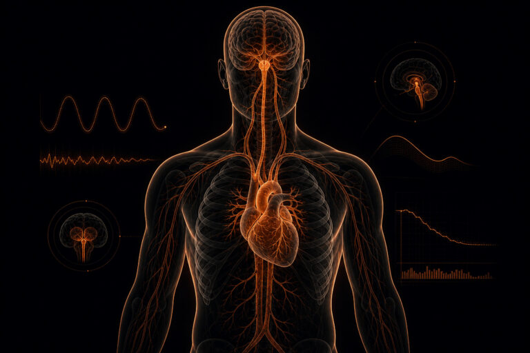
The vagus nerve (cranial nerve X) is the longest cranial nerve in the body, originating in the dorsal vagal complex and nucleus ambiguus of the medulla oblongata and extending through the thorax and into the abdomen. Its efferent cardiac branch innervates the sinoatrial node, where acetylcholine release from vagal terminals slows pacemaker firing and reduces heart rate. The reach of the nerve is broad, but the cardiac branch is what drives the HRV signals most researchers and clinicians are working with day to day.6
The nerve carries two classes of efferent fibers relevant to autonomic function. Myelinated B-fibers project from the nucleus ambiguus to the sinoatrial and atrioventricular nodes; these are the fibers whose activity is most directly reflected in HRV. Unmyelinated C-fibers project from the dorsal vagal nucleus to the viscera, regulating digestion and inflammatory responses. The key distinction is speed: the myelinated branch responds rapidly enough to generate the beat-to-beat variability that HRV metrics capture, while the unmyelinated branch operates on slower, less rhythmically quantifiable timescales and does not contribute meaningfully to standard HRV indices.6
The connection to HRV is mechanistic, not merely correlational. Efferent vagal firing slows the SA node; withdrawal of that firing speeds it back up. The oscillation between vagal braking and release, synchronized with respiratory cycles, produces respiratory sinus arrhythmia, the physiological substrate of HF HRV and RMSSD. This beat-to-beat entrainment is precisely why RMSSD is considered the most direct non-invasive index of cardiac vagal activity.4
How Vagal Tone Is Estimated: HRV as a Proxy
No ambulatory method measures vagal tone directly. Research protocols estimate it from the HRV indices most sensitive to vagal modulation: RMSSD, pNN50, and HF power. The 1996 Task Force of the European Society of Cardiology established the methodological standards for these measurements, including the requirement for at minimum 5-minute stationary recordings for short-term frequency-domain analysis, or 24-hour recordings for time-domain norms. Those recording requirements exist for a reason: the signal is sensitive to posture, breathing rate, and motion in ways that can easily swamp the autonomic information you are trying to extract.4
ECG-derived RR intervals, the time between successive R-peaks in the cardiac waveform, remain the reference standard for HRV analysis. PPG-derived interbeat intervals, captured optically by wearable sensors using photoplethysmography, provide an alternative in research and monitoring workflows where ECG electrode placement is impractical. PPG-derived intervals correspond to the peak-to-peak interval of the optical pulse waveform and introduce additional sources of variability absent from ECG, including motion artifact and peripheral vascular resistance changes.1
Research validating PPG-derived HRV against ECG-derived HRV generally shows acceptable agreement for RMSSD under controlled, low-motion conditions, with wider agreement limits during movement. That said, acceptable group-level agreement does not guarantee individual-level precision, which is a distinction worth keeping in mind when interpreting a single person’s data. Clinicians and researchers using optically derived interbeat intervals should note the distinction and apply appropriate interpretive caution. PPG-based HRV supports population-level trend analysis and longitudinal monitoring well; it is not a direct substitute for ECG HRV in diagnostic contexts.3
What High and Low Vagal Tone Indicate in Research Literature
Across a large body of physiological research, higher vagally mediated HRV, used as a proxy for vagal tone, has been associated with more effective parasympathetic regulation of heart rate, better emotional regulation capacity, and greater cardiovascular resilience under stress. Lower vagally mediated HRV has been associated with increased stress load, poorer autonomic recovery after exertion, and adverse cardiometabolic profiles in population studies. These are consistent findings across many research groups, which is part of why vagal tone has attracted so much clinical and commercial attention in recent years.1
What changes if you look more carefully is the precision of those claims. These are population-level statistical associations derived from group comparisons, not diagnostic thresholds for individual assessment. A single individual’s RMSSD or HF HRV value does not diagnose autonomic dysfunction. Reference values vary substantially by age (vagally mediated HRV declines with aging), sex (women show higher HF HRV in some age bands), fitness status, posture, time of day, and recording conditions, all factors the 1996 Task Force guidelines require researchers to standardize and report before drawing conclusions.4
Thayer and Lane’s neurovisceral integration model offers a theoretical framework for why vagal tone is relevant beyond cardiac regulation: the prefrontal-subcortical circuits governing inhibitory control of the heart also modulate cognitive and emotional responses, suggesting that vagal tone reflects a broader index of self-regulatory capacity.5 The harder question is how far that framework actually extends. Grossman (2023) has raised substantive critiques of both the construct validity of “vagal tone” as commonly used and the autonomic specificity assumptions embedded in polyvagal theory: evidence, accuracy, and clinical use, warranting careful framing of any causal claims about what HRV actually tells you about the nervous system.2
Factors That Influence Vagal Tone: What the Evidence Shows
Several modifiable factors have been associated with higher vagally mediated HRV in controlled studies, though the strength and durability of each association varies across research designs. Aerobic exercise training shows the most consistent evidence: longitudinal studies find that regular endurance training is associated with resting HF HRV increases, particularly in previously sedentary adults. The effect appears dose-related in some studies, suggesting that training load matters, not just the fact of exercising at all.1 Controlled breathing practices, specifically slow-paced breathing near the resonance frequency (approximately 0.1 Hz, or roughly 6 breaths per minute), have been associated with increased RSA amplitude in short-term protocols, though the durability of these effects outside experimental conditions is less established and the mechanisms underlying any long-term shift remain an open question.3
Sleep quality is an emerging area of investigation. Studies linking poor sleep to suppressed HRV suggest a bidirectional relationship between autonomic regulation and sleep architecture, though causal direction remains under investigation and the field has not yet produced consensus on mechanism. The suppression appears across multiple HRV indices, not just RMSSD, which suggests the relationship is not a measurement artifact. Cold facial exposure produces acute, transient vagal responses via the diving reflex, a well-characterized physiological effect; this is mechanistically real but does not constitute a sustained intervention, and the evidence for lasting vagal tone changes from cold exposure alone is thin.5
Non-modifiable factors shape the baseline regardless of behavior. Age is the strongest: vagally mediated HRV declines progressively across the lifespan, with the steepest decline in middle age. Sex differences are present, particularly in younger cohorts, with females showing higher HF HRV in some studies, though these differences narrow with age and fitness status. Athletic training status introduces substantial confounding in any cross-sectional comparison, which is why comparisons between athletes and non-athletes require careful control for fitness level and training history. Interpreting an individual’s vagal tone estimate without accounting for these contextual factors produces conclusions that are, at best, misleading.4
Vagal Tone, HRV, and Continuous Monitoring: Clinical and Research Context
Single-point HRV snapshots capture vagal tone at one moment under one set of conditions. That matters because conditions change constantly, and a single reading carries no information about direction of travel. Researchers increasingly argue that longitudinal, multi-day recordings provide far more informative data about autonomic regulation trends than any isolated measurement. Tracking how vagally mediated HRV shifts across recovery periods, stress exposure, sleep cycles, and training loads reveals patterns that a resting morning measurement simply misses.1
Wearable optical sensors have made longer-duration HRV data collection feasible in research and monitoring workflows. Continuous PPG recording generates interbeat interval time series from which RMSSD and HF HRV can be computed across naturalistic conditions, including sleep, light activity, and transitions between them. The interpretive challenge is that ambulatory recordings introduce noise sources that are absent from supervised lab protocols. Motion artifact, posture changes, and variable skin contact all affect signal quality in ways a stationary recording avoids. Validated algorithms and appropriate artifact rejection are required before drawing autonomic conclusions from wearable data; applying lab-derived norms to noisy ambulatory signals without adjustment is a common and consequential error.3
In clinical workflow terms, Remote Therapeutic Monitoring (RTM; CPT codes 98975 to 98981) provides a billing and supervision framework for clinicians to oversee remote data collection and engage therapeutically with patients between visits. RTM is distinct from Remote Patient Monitoring (RPM; CPT codes 99453 to 99458), which applies to physiological data collection for chronic condition management under different coverage rules. Sensor Bio devices are not FDA-cleared medical devices; they are designed to support RTM workflows under clinician oversight, providing longitudinal autonomic signal data for research and therapeutic monitoring purposes, not to diagnose or treat autonomic disorders.4 Clinicians evaluating any wearable-derived HRV data for individual patient decisions should apply standard autonomic physiology principles and account for the signal limitations of optically derived interbeat intervals.
| HRV Index | Domain | Vagal Sensitivity | Recording Requirement | Signal Source |
|---|---|---|---|---|
| RMSSD | Time | High (primary vagal index) | ≥5 min stationary | ECG or validated PPG |
| pNN50 | Time | High | ≥5 min stationary | ECG or validated PPG |
| HF power (0.15–0.4 Hz) | Frequency | High (RSA band) | ≥5 min stationary; controlled breathing preferred | ECG or validated PPG |
| SDNN | Time | Mixed (vagal + sympathetic) | 24 h preferred for full norms | ECG reference standard |
| LF/HF ratio | Frequency | Low (contested interpretation) | ≥5 min stationary | ECG; PPG with artifact caution |
PPG vs. ECG for Estimating Vagal Tone: Trade-offs
ECG-derived RR intervals are the reference standard for HRV research. The R-peak is a sharp, high-amplitude electrical event that can be detected with sub-millisecond precision, making ECG-derived interbeat intervals the most accurate input for vagal tone estimation. Frequency-domain analysis and time-domain metrics computed from ECG RR intervals reflect cardiac vagal modulation with minimal ambiguity about signal origin. When precision matters most, ECG is the appropriate tool, and that standard should remain visible even when practical constraints push workflows toward optical alternatives.4
PPG-derived interbeat intervals, measured optically at the wrist or finger, introduce several sources of additional variance. The PPG waveform peak is broader and more susceptible to motion artifact, peripheral vasoconstriction, and skin perfusion changes. Pulse transit time, the delay between cardiac ejection and peripheral pulse arrival, introduces a systematic offset relative to the true RR interval; this offset is relatively stable at rest but varies with blood pressure changes and physical activity. Studies comparing PPG-derived and ECG-derived RMSSD under controlled, resting conditions generally find acceptable agreement at the group level; individual-level agreement is wider and degrades with motion.1
For longitudinal research applications, tracking autonomic trends across days or weeks, wrist PPG provides a practical trade-off: lower per-reading precision offset by the ability to obtain far more data points than periodic ECG recording allows. The interpretive frame must shift accordingly. PPG-derived HRV data supports trend detection and population-level analysis more reliably than single-session diagnostic precision, and using it well means understanding where that boundary sits and designing your interpretation around it rather than ignoring the constraint.3
Limits and Pitfalls When Interpreting Vagal Tone Estimates
Several interpretive errors recur when vagal tone estimates move from research papers into applied contexts. The first is treating RMSSD or HF HRV as a direct measurement of vagal tone rather than a proxy. Vagal tone is a construct; the measured signal is influenced by vagal activity but also by sympathetic tone, respiratory rate, baroreflex gain, and recording methodology. A change in RMSSD tells you something shifted in the autonomic system. It does not tell you precisely what shifted, or why.2
The second pitfall is applying group-level reference ranges to individual interpretation. Published HRV norms are derived from population samples and carry wide inter-individual variability. An individual’s “normal” is best understood relative to their own baseline trend, not relative to population percentiles drawn from a different demographic with different age, sex, and fitness distributions. Using a published normative table to label someone’s RMSSD as “low” without accounting for those factors is a category error that generates more noise than insight.3
Third, recording conditions matter enough to invalidate comparisons made without standardization. Supine versus orthostatic posture changes HF HRV substantially. Respiratory rate modulates RSA amplitude; paced breathing protocols produce systematically different results than spontaneous breathing. Time of day, recent meal, caffeine intake, and acute psychological state all influence HRV readings in measurable ways. Two recordings from the same person taken under different conditions may not be meaningfully comparable, even when expressed in the same units.4
Finally, Grossman’s (2023) critique of autonomic specificity assumptions deserves serious consideration. The implicit claim embedded in much vagal tone research is that HRV straightforwardly indexes vagal activity. That claim relies on assumptions about the independence of sympathetic and parasympathetic branches that the evidence only partially supports. Researchers working with HRV data should remain cautious about strong causal language connecting HRV changes to specific autonomic mechanisms, and should treat effect-size claims in the vagal tone literature with the skepticism that any contested construct warrants.2
FAQ
What does vagal tone mean in physiological terms?
Vagal tone refers to the baseline level of tonic parasympathetic outflow from the vagus nerve (cranial nerve X) to the heart and visceral organs. Higher tonic vagal activity slows the sinoatrial node and increases beat-to-beat heart rate variability. Because vagal tone cannot be observed directly in ambulatory settings, researchers estimate it using vagally mediated HRV indices, most commonly RMSSD and HF power, derived from ECG or validated PPG recordings.3
Is vagal tone the same as HRV?
No. Vagal tone is the underlying physiological construct: the tonic efferent activity of the vagus nerve. HRV, specifically vagally mediated indices like RMSSD and HF HRV, is a proxy signal used to estimate that construct. HRV is also shaped by sympathetic activity, respiratory rate, baroreflex function, and recording conditions. Treating the two as synonymous overstates what HRV measurements can claim about autonomic state.2
What causes low vagal tone according to the research literature?
In research populations, lower vagally mediated HRV has been associated with chronic psychological stress, poor sleep quality, sedentary behavior, advancing age, and certain cardiometabolic conditions. These are population-level associations from physiological studies, not diagnostic criteria. Individual HRV values vary substantially based on age, sex, fitness level, posture, time of day, and recording methodology; low HRV relative to population norms does not constitute a diagnosis.1
Can vagal tone be changed over time?
Research evidence suggests aerobic exercise training and slow-paced breathing practices are associated with higher vagally mediated HRV in study populations, consistent with an increase in tonic vagal activity. Evidence quality varies by intervention, duration, and population. These findings come from controlled research studies; individual responses are context-dependent. The research framing matters: associations in study populations do not guarantee equivalent effects for any specific individual.1
How is vagal tone measured in wearable research settings?
Vagal tone cannot be directly measured by wearable sensors. Research protocols use HRV indices computed from interbeat interval time series derived either from ECG or from validated PPG optical sensors. RMSSD, pNN50, and HF power are the indices most sensitive to vagal modulation. PPG-based optical sensors capture interbeat intervals optically rather than electrically, introducing additional variance from motion and peripheral perfusion; ECG-derived RR intervals remain the reference standard for vagal tone estimation.4
What is the difference between vagal tone and polyvagal theory?
Vagal tone is a physiological construct describing the tonic efferent activity of the vagus nerve, estimated via HRV indices. Polyvagal theory (Porges, 2011) is a broader theoretical framework proposing that the vagus nerve has two distinct functional branches, the myelinated ventral vagal complex and the unmyelinated dorsal vagal complex, with separate behavioral and social roles. The theory is influential in trauma-informed care contexts but has received substantive critique in the autonomic neuroscience literature regarding its anatomical and physiological assumptions. For a detailed examination of those critiques and the current state of the evidence, see polyvagal theory: evidence, accuracy, and clinical use.26
Why does the vagus nerve affect heart rate variability?
The vagus nerve’s myelinated cardiac efferent fibers innervate the sinoatrial node, the heart’s primary pacemaker. Increased vagal firing releases acetylcholine at the SA node, slowing its spontaneous depolarization rate and reducing heart rate. Decreased vagal firing allows the rate to accelerate. The respiratory-driven oscillation of this vagal brake, speeding with inhalation and slowing with exhalation, produces respiratory sinus arrhythmia, the physiological basis of RMSSD and HF HRV. This beat-to-beat modulation is why vagally mediated HRV indices are the primary non-invasive proxy for cardiac vagal tone.5
References
Reference ranges and population norms for vagal tone proxies
Population norms for RMSSD vary substantially by age, sex, recording conditions, and population health status. No single threshold defines adequate vagal tone across all adults. 7
Nunan et al. (2010) provides the most widely cited normative dataset, drawn from a quantitative systematic review of short-term HRV values in 1,066 healthy adults measured under standardized resting conditions. Adults aged 20–29 showed mean RMSSD values of approximately 42–68 ms in supine short-term recordings. Values decline progressively across age decades; mean RMSSD in adults aged 60 and above falls substantially below values observed in the youngest cohort. 7
Sex differences are clinically relevant. Before menopause, females generally show higher HF power and comparable or higher RMSSD than age-matched males, a pattern consistent with hormonal modulation of autonomic activity. This difference narrows after menopause. Stratified reference values accounting for both age and sex provide more interpretable context than age-only norms.
Measurement conditions introduce systematic variability that can exceed many reported group differences. Supine recordings yield higher RMSSD than standing by approximately 10–20 ms in typical adults. Nocturnal values consistently exceed daytime values. Short-term 5-minute standardized recordings differ meaningfully from 24-hour ambulatory recordings, and norms derived from one protocol do not apply to the other. Any cross-sectional comparison to population percentiles must account for the recording conditions under which the reference data were collected. 4
References
- Laborde S, Mosley E, Mertgen A, Sauter C, Engel M, Raab M. (2024). Vagally mediated heart rate variability: A meta-analytic review of associations with psychological and physical health. Clinical Physiology and Functional Imaging. https://doi.org/10.1111/cpf.12888
- Grossman P. (2023). The myth of autonomic specificity: Issues with the concept of vagal tone and its measurement. Biological Psychology, 177, 108470. https://doi.org/10.1016/j.biopsycho.2023.108470
- Shaffer F, Ginsberg JP. (2017). An overview of heart rate variability metrics and norms. Frontiers in Public Health, 5, 258. https://doi.org/10.3389/fpubh.2017.00258
- Task Force of the European Society of Cardiology and the North American Society of Pacing and Electrophysiology. (1996). Heart rate variability: Standards of measurement, physiological interpretation, and clinical use. Circulation, 93(5), 1043–1065. https://doi.org/10.1161/01. CIR.93.5.1043
- Thayer JF, Lane RD. (2009). Claude Bernard and the heart–brain connection: Further elaboration of a model of neurovisceral integration. Neuroscience & Biobehavioral Reviews, 33(2), 81–88. https://doi.org/10.1016/j.neubiorev.2008.08.004
- Porges SW. (2011). The Polyvagal Theory: Neurophysiological Foundations of Emotions, Attachment, Communication, and Self-Regulation. W. W. Norton & Company.
- Nunan D, Sandercock GRH, Brodie DA. A quantitative systematic review of normal values for short-term heart rate variability in healthy adults. Annals of Noninvasive Electrocardiology. 2010;15(4):301–309. PubMed: 20552350.