

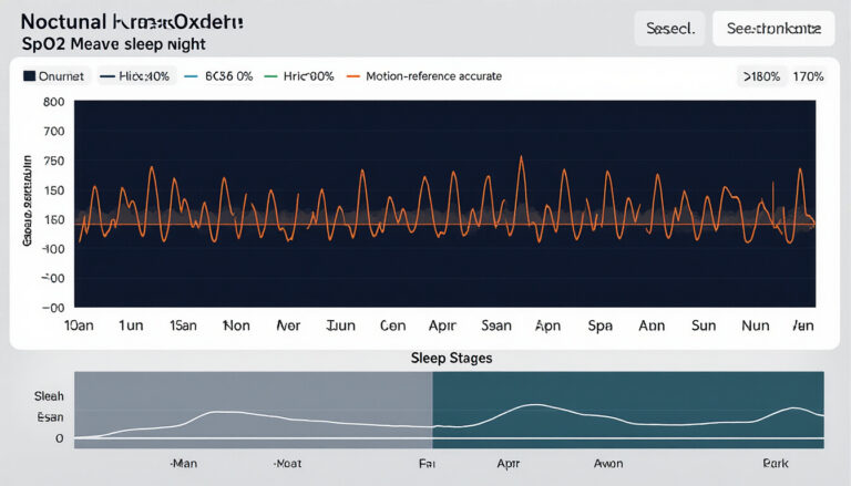
SpO2 measured at night is generally reliable under stable conditions, but nocturnal accuracy depends on signal quality, device placement, sleep position, and the specific hardware and algorithm producing the value. Three factors explain how to evaluate that reliability: the physics of pulse oximetry, how sleep physiology alters the measurement environment, and where reflectance-based wearables diverge from clinical transmission oximeters in error profile.
is spo2 accurate at night depends on the sensor, fit, perfusion, motion, skin tone validation, and whether drops repeat across nights instead of appearing as isolated artifacts.
is spo2 accurate at night validation checklist
This short retrofit section clarifies the exact search intent for is spo2 accurate at night while preserving the original article and adding practical verification points for readers.
Verification checklist
- Look for repeated overnight patterns rather than one sudden single-sample drop.
- Check fit, motion, cold hands, nail polish, and perfusion as artifact sources.
- Seek clinical testing for persistent low readings or symptoms instead of relying on a consumer wearable alone.
Related Sensor Bio reading
Authoritative references
What SpO2 is and how pulse oximetry measures it
SpO2 is the percentage of hemoglobin in arterial blood that is bound to oxygen. Pulse oximetry estimates SpO2 non-invasively by applying the Beer-Lambert law: two wavelengths of light (typically 660 nm red and 940 nm infrared) are transmitted through or reflected from perfused tissue, and the differential absorption ratio between oxyhemoglobin and deoxyhemoglobin yields the saturation estimate.1
The underlying signal is a photoplethysmography (PPG) waveform, the oscillating pattern of light absorption synchronized to the cardiac cycle. The pulsatile (AC) component of the PPG waveform corresponds to arterial blood volume changes; the ratio of AC to DC amplitude at each wavelength, known as the R-ratio, is the direct input to the SpO2 algorithm.2 Clinical transmission oximeters place a light emitter and detector on opposite sides of a fingertip. Reflectance devices (rings, wrist bands) use a co-located emitter and detector, capturing backscattered light. Both modes depend on adequate pulsatile signal amplitude, quantified by the perfusion index (PI), which expresses the AC-to-DC amplitude ratio as a percentage.
When PI falls below approximately 0.3%, clinical oximeters issue reliability warnings.1 Consumer reflectance devices, by contrast, typically do not expose the perfusion index to end users at all, which matters directly for interpreting nocturnal SpO2 data. The full PPG signal chain, from photon emission through waveform extraction to saturation calculation, is what explains why nighttime conditions systematically degrade measurement quality.
How nighttime physiology changes the measurement environment
Sleep imposes systematic changes on peripheral circulation and respiratory pattern that directly affect SpO2 measurement quality. During non-REM sleep, parasympathetic dominance promotes peripheral vasoconstriction as core body temperature drops and blood redistributes toward central compartments. This vasoconstriction reduces pulsatile amplitude at the finger and wrist, lowering the perfusion index and degrading signal quality even without movement.1
REM sleep introduces sympathetic surges that cause transient increases in heart rate and respiratory irregularity. Normal respiratory variations during REM, including brief pauses in ventilation at sleep stage transitions, produce transient SpO2 dips of 2–4% that are physiological. Slow-wave sleep (N3) is associated with the lowest metabolic demand and most stable respiratory pattern; SpO2 values during N3 are typically the most consistent in healthy adults.5
Nocturnal SpO2 accuracy fluctuates with sleep stage, body position changes, ambient temperature, and individual vascular tone, never holding steady across the full night. A recording that appears meaningful as a summary statistic may contain stretches of low-confidence data that inflate or suppress the apparent oxygen desaturation index. Point-in-time SpO2 values are unreliable without accompanying signal quality metadata.
Device and signal factors that degrade nocturnal SpO2 accuracy
Motion artifact, contact mechanics, low perfusion index, and melanin-related optical bias are the four primary factors that degrade SpO2 accuracy during sleep.
Motion artifact. Even low-amplitude sleep movements, such as repositioning a hand or rolling to one side, introduce mechanical disturbances into the PPG waveform that corrupt the R-ratio calculation. Clinical oximeters apply motion-resistant signal averaging; consumer reflectance devices vary widely in their artifact-rejection capability.4
Contact pressure and sleep position. Prone and lateral positions apply compression to the measurement site, generating venous pulsation that the oximeter cannot distinguish from arterial pulsation. This venous artifact produces artificially low SpO2 estimates. The effect is more pronounced in ring- and wrist-form devices than in forehead sensors because extremity vessels are more susceptible to extrinsic compression.12
Low perfusion index. Cold ambient temperatures, peripheral vascular disease, or vasoconstriction from sleep cause PI to fall. Below the 0.3% threshold, SpO2 readings become unreliable; accuracy improves progressively with signal amplitude above that floor.
Melanin-related optical bias. Melanin absorbs light across the wavelengths used by pulse oximetry, reducing signal-to-noise ratio and introducing a systematic positive bias in individuals with darker skin tones. A 2020 NEJM analysis of 10,789 patients found that Black patients had three times the frequency of occult hypoxemia undetected by pulse oximetry compared to white patients.3 The FDA issued a safety communication in 2021 acknowledging this limitation and calling for improved device testing across skin tones.7
What the evidence says about nocturnal SpO2 accuracy in practice
Clinical transmission oximeters validated under ISO 80601-2-61 must achieve root-mean-square accuracy of ±2% ARMS across the 70–100% SpO2 range under controlled testing conditions.6 Reflectance wearables show wider error distributions: a systematic review of wearable cardiac monitoring found mean absolute errors of 2–5% for reflectance SpO2 during sleep compared to co-oximetry reference, with larger errors at lower saturation values and in subjects with higher melanin levels.4
| Device type | SpO2 range tested | Mean error vs. reference | Study design | Citation |
|---|---|---|---|---|
| Transmission pulse oximeter (clinical) | 70–100% | ±2% ARMS (standard) | ISO standard testing | 6 |
| Reflectance wearable (ring, wrist) | 80–100% | 2–5% mean absolute error | Bench and clinical validation | 4 |
| Clinical oximeter (darker skin tones) | 88–100% | +3–4% positive bias | Retrospective cohort (n=10,789) | 3 |
| Overnight continuous oximetry (sleep apnea screening) | Full night | Sensitivity ~75% for AHI ≥15 | Prospective screening study | 8 |
In sleep apnea screening, the oxygen desaturation index (ODI) is the clinically relevant aggregate metric, counting events per hour where SpO2 drops by ≥3% or ≥4%. The 3% ODI carries moderate sensitivity (approximately 65–80%) for detecting moderate-to-severe obstructive sleep apnea, but specificity is lower, meaning false positives occur.8 A negative overnight oximetry result does not rule out sleep apnea; diagnostic polysomnography or home sleep testing remains required for clinical confirmation.5
When to trust and when not to trust a nocturnal SpO2 reading
Three indicators distinguish trustworthy nocturnal SpO2 data from artifact: a perfusion index above 0.3% throughout the recording, signal quality flags showing acceptable waveform amplitude, and a sample rate of at least 1 Hz for ODI calculation.5 A summary SpO2 value without those indicators conveys false precision.
Clinical-grade diagnostic sleep studies require type III or type IV portable monitoring devices or in-lab polysomnography, not consumer wearables. Wearable ring and wrist devices provide screening-level data that can motivate further evaluation but cannot substitute for a diagnostic study. Research-grade platforms that preserve raw PPG waveform data enable post-hoc signal quality assessment and artifact rejection that consumer devices, which discard waveform data after deriving the SpO2 estimate, cannot support.24
Data are likely uninterpretable when the device was compressed under the body for an extended period, when ambient temperature caused significant peripheral vasoconstriction, or when the device was displaced from the measurement site. Sustained SpO2 below 90% for more than five consecutive minutes is the pattern associated with confirmed sleep-disordered breathing; isolated low readings without that sustained pattern point more often to measurement artifact than a true respiratory event.5
For clinicians and researchers evaluating nocturnal biosignal data at scale, access to continuous raw PPG waveform output transforms SpO2 from a black-box estimate into an auditable, reanalyzable data stream. The underlying measurement physics are covered in our photoplethysmography fundamentals guide; signal quality methodology is detailed on the Sensor Bio science page. To explore research and clinical platform options, visit the get started page.
Frequently asked questions about nocturnal SpO2 accuracy
Does SpO2 naturally drop at night in healthy people?
Transient SpO2 dips of 2–4% occur in healthy adults during REM sleep, reflecting normal autonomic and respiratory changes at sleep stage transitions. Brief as they are, these fluctuations are physiological, not a sign of pathology. Sustained SpO2 below 90% lasting more than five consecutive minutes is not a pattern typically seen in otherwise healthy adults and is associated with sleep-disordered breathing in clinical populations. Single low readings without contextual signal quality data should be interpreted with caution: they more commonly reflect measurement artifact than a true respiratory event.5
How accurate is a wearable ring or wrist device for overnight SpO2?
Reflectance-mode wearables show mean absolute errors of approximately 2–5% compared to co-oximetry reference under ideal conditions, with larger errors under motion, low perfusion index, and in individuals with higher melanin levels. ISO 80601-2-61 sets ±2% ARMS as the minimum accuracy standard for clinical spot-check oximeters, tested across the 70–100% saturation range with controlled hypoxemia protocols.6 Most consumer wearables are validated on narrower demographic samples and may not meet that standard across all skin tones or physiological states encountered during sleep.34
Can I use overnight SpO2 data to screen for sleep apnea?
Overnight pulse oximetry can calculate the oxygen desaturation index (ODI), counting events per hour where SpO2 drops by ≥3% or ≥4%. The 3% ODI has moderate sensitivity (approximately 65–80%) for detecting moderate-to-severe obstructive sleep apnea in appropriately selected populations, with lower specificity.8 Even a negative wearable result cannot rule out sleep apnea, and a positive finding still requires confirmation with a diagnostic polysomnogram or home sleep test meeting clinical standards. Wearable data is screening-level evidence, not a diagnostic output, and should be treated accordingly when communicating findings to patients or study participants.5
Why does sleeping position affect SpO2 accuracy?
Prone and lateral positions apply compression to the finger or wrist, generating venous pulsation that pulse oximeters cannot distinguish from arterial pulsation. This venous artifact shifts the R-ratio toward values that produce falsely low SpO2 estimates. The magnitude of the effect depends on the pressure applied and the compliance of the underlying vessels. Clinical oximeters flag perfusion index below 0.3% as a signal quality warning. Most consumer devices do not expose this indicator to users, making position-related artifact difficult to detect without access to the underlying PPG waveform for post-hoc review.12
Does skin tone affect nighttime SpO2 accuracy?
Yes. Melanin absorbs light across the wavelengths used by pulse oximeters, reducing signal quality and introducing a systematic positive bias in individuals with darker skin tones: the device reads higher than the true arterial saturation. A 2020 analysis published in the New England Journal of Medicine found that Black patients had three times the rate of occult hypoxemia missed by pulse oximetry compared to white patients, across a cohort of 10,789 subjects.3 The FDA acknowledged this limitation in a 2021 safety communication applicable to all pulse oximeter devices, both clinical and consumer-grade, and called for more inclusive device testing protocols.7
Is a single low overnight SpO2 reading cause for concern?
An isolated low SpO2 reading, for example a single data point at 88% on a wearable during sleep, most commonly reflects signal artifact from movement or probe displacement rather than true physiological desaturation. Clinically meaningful nocturnal hypoxemia is characterized by sustained events: SpO2 below 90% for more than five consecutive minutes, or an ODI of five or more events per hour in the appropriate clinical context, per AASM scoring standards.5 Single-point readings without accompanying signal quality data are insufficient for clinical interpretation. If sustained low readings appear across multiple nights, evaluation by a sleep medicine clinician is the appropriate next step.
References
- Jubran A. Pulse oximetry. Critical Care. 1999;3(2):R11–R17. PMID: 11094477.
- Allen J. Photoplethysmography and its application in clinical physiological measurement. Physiological Measurement. 2007;28(3):R1–R39. PMID: 17322588.
- Sjoding MW, Dickson RP, Iwashyna TJ, Gay SE, Valley TS. Racial bias in pulse oximetry measurement. New England Journal of Medicine. 2020;383(25):2477–2478. PMID: 33326721.
- Castaneda D, Esparza A, Ghamari M, Soltanpur C, Nazeran H. A review on wearable technology for cardiac monitoring: taxonomy, trends, opportunities and challenges. Sensors. 2020;19(9):1966. PMID: 31035408.
- Berry RB, Brooks R, Gamaldo C, et al. The AASM Manual for the Scoring of Sleep and Associated Events. Version 2.4. American Academy of Sleep Medicine; 2017.
- ISO 80601-2-61:2017. Medical electrical equipment, Part 2-61: Particular requirements for basic safety and essential performance of pulse oximeter equipment. International Organization for Standardization; 2017.
- U. S. Food and Drug Administration. Pulse oximeter accuracy and limitations: FDA safety communication. February 19, 2021.
- Lévy P, Kohler M, McNicholas WT, et al. Obstructive sleep apnoea syndrome. Nature Reviews Disease Primers. 2015;1:15015. PMID: 27188535.
Is SpO2 accurate at night? How wrist and ring PPG sensors work
The core question — is SpO2 accurate at night when measured by a wrist or ring device? — begins with understanding the physics. Consumer wearables use photoplethysmography: green LEDs for heart rate, and red plus infrared LEDs for blood oxygen. The ratio of absorbed light at those two wavelengths changes as hemoglobin picks up or releases oxygen. Firmware converts that ratio into an SpO2 percentage using a calibration curve built from controlled lab studies. The precision of that calibration — and who participated in it — sets the ceiling on overnight accuracy before environmental factors intervene.
Ring-form sensors (Oura, Samsung Galaxy Ring) tend to outperform wrist bands for SpO2 because the finger has higher skin perfusion, thinner tissue overlying the capillary bed, and a more stable contact surface during sleep. Wrist devices contend with radial-artery depth variability, hair follicles, tattoo pigment, and the strap tension that shifts whenever a sleeper changes position. Neither form factor has FDA clearance as a diagnostic oximeter; both target ±2–3% accuracy under ideal lab conditions, which widens meaningfully in real overnight use.
Motion and perfusion artifacts: the main accuracy killers
Motion artifact is the largest single source of error in overnight SpO2 data. When a sleeper turns over, the sudden limb movement generates a PPG signal that the algorithm may briefly misread as a low-oxygen event. Most modern devices apply signal-quality scoring and drop or flag corrupt windows, but short motion artifacts — under five to ten seconds — often slip through as apparent SpO2 dips. Before interpreting any overnight low reading, checking the device’s accelerometer log or signal-quality indicator is the first diagnostic step.
Peripheral perfusion drops during sleep in cold rooms or in people with Raynaud’s phenomenon, poor circulation, or low cardiac output. When capillary blood flow at the wrist or finger diminishes, the PPG signal amplitude falls, making it harder for the sensor to distinguish the arterial pulse from background noise. The result is often a falsely low SpO2 or a dropped reading. Warming the room, using a fitted ring rather than a wrist band, and ensuring the device is snug without being circulation-restricting all measurably improve overnight data quality.
Fit, skin tone, and sensor placement considerations
The question of whether is SpO2 accurate at night also depends on the device fit and, importantly, on skin tone. Darker skin tones absorb more red and infrared light, which can shift the apparent hemoglobin ratio the sensor reads, causing the algorithm to overestimate SpO2 — meaning a device may report 95% when true saturation is 90–92%. The FDA flagged this issue in a 2021 safety communication, and several academic groups have since quantified the bias in FDA-cleared clinical pulse oximeters, where the problem is well-documented. Consumer wearables, with less rigorous calibration diversity, are subject to at least the same risk.
Proper fit matters beyond just comfort. A ring or band that rotates freely on the finger or wrist repositions the LED over a different skin region between measurements, breaking the consistency the algorithm assumes. Sizing guides from manufacturers typically recommend the loosest size that still holds the sensor flat against the skin. If the device slides noticeably when you shake your wrist gently, it is too loose for reliable overnight SpO2 data.
What overnight desaturation patterns can and cannot tell you
Is SpO2 accurate at night in the sense of revealing a medically meaningful pattern? For trend detection — yes, with caveats. A wearable that consistently shows SpO2 spending more than 10–15 minutes below 90% across multiple nights is flagging something worth investigating, even accounting for device error. Clinicians use a metric called T90 (time below 90% saturation) and an oxygen desaturation index (ODI, desaturation events per hour) as sleep-disordered-breathing markers; consumer devices approximate these but do not report them in standardized clinical units.
What overnight wearable SpO2 cannot tell you: the cause of the desaturation. Central apnea, obstructive apnea, hypoventilation, obesity hypoventilation syndrome, altitude-related changes, and cardiac-output reduction can all produce overnight SpO2 dips. Distinguishing them requires simultaneous airflow measurement, respiratory-effort belts, end-tidal CO2, and EEG staging — none of which consumer wearables provide. A repeated SpO2 pattern is a prompt to seek evaluation, not a diagnosis of any specific condition.
When to seek medical-grade testing for sleep apnea
The decision to move from wearable data to clinical evaluation does not require certainty about whether is SpO2 accurate at night on your specific device. It requires recognizing that the stakes of missing sleep-disordered breathing are high enough to warrant proper testing. Home sleep tests use FDA-cleared finger-clip pulse oximeters accurate to ±1–2% and add nasal airflow thermistors and respiratory-effort belts. In-lab polysomnography adds EEG, EOG, and EMG channels for full staging and arousal scoring. Both provide orders of magnitude more information than a wrist or ring sensor, making them the definitive answer when is SpO2 accurate at night becomes a clinically important question.
Specific prompts to seek evaluation: consistent overnight SpO2 below 90%, waking unrefreshed despite adequate sleep opportunity, witnessed apneas reported by a bed partner, morning headaches attributable to CO2 retention, excessive daytime sleepiness on the Epworth scale, or any combination of loud snoring and the above. Wearable SpO2 data can usefully accompany a clinical appointment — as trend documentation, not as a diagnostic report — and can help quantify symptom burden over weeks rather than the single night captured in a clinical study.