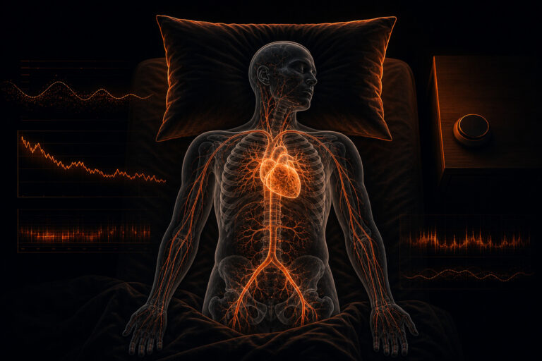
A low heart rate during sleep is normal in most adults. The heart rate typically falls 10 to 30 percent below the daytime resting rate during non-REM sleep as parasympathetic nervous system activity increases.1 That drop is not a malfunction. It is the body doing exactly what it should during recovery.
For most people, nocturnal heart rates between 40 and 60 beats per minute (bpm) reflect healthy autonomic regulation rather than pathology. The clinical picture changes when rates drop below 40 bpm repeatedly, when the pattern is accompanied by symptoms such as syncope or palpitations on waking, or when the slowing correlates with apneic events. Understanding the physiology behind nocturnal cardiac slowing helps you distinguish normal sleep adaptation from patterns that merit clinical evaluation. This article covers the mechanism, normal reference ranges, concerning patterns, recovery relevance, and measurement trade-offs for low heart rate during sleep, drawing on published human studies throughout.
What is sleep bradycardia
Bradycardia is defined as a heart rate below 60 bpm. During sleep, this threshold is routinely crossed by healthy adults without pathological consequence. The physiological driver is parasympathetic dominance: as wakefulness gives way to non-REM sleep, the autonomic nervous system shifts toward vagal activity, which slows sinoatrial node firing and reduces cardiac output to match the body’s lowered metabolic demand.1 This shift is not subtle. During slow-wave sleep (N3 stage), heart rates of 40 to 55 bpm are commonly recorded in otherwise healthy individuals.2 The electrocardiographic and optical biosignal signatures of this transition are well characterized in the literature on sympathetic vs parasympathetic nervous system activity.
The term “sleep bradycardia” describes this physiologically appropriate slowing and carries no inherent clinical severity. Athletes and highly conditioned individuals often show resting nocturnal rates in the mid-30s, the result of chronically elevated vagal tone and cardiac adaptation to endurance training, not a sign of disease. Low heart rate during sleep driven by vagal up-regulation is mechanistically distinct from pathological bradycardia caused by conduction system disease or pharmacological suppression of sinoatrial automaticity. Appreciating that distinction is the starting point for interpreting any nocturnal heart rate value. Context determines whether a slow heart rate at night is a biomarker of good recovery or a signal that warrants further evaluation.
Normal nocturnal heart rate: what the data show
Published population data indicate that average nocturnal heart rates in healthy adults fall between 50 and 65 bpm, with individual ranges extending from the upper 30s to the low 70s depending on age, fitness level, and sleep stage.3 A quantitative systematic review by Nunan et al. (2010) documented normative ranges for short-term heart rate variability (HRV) metrics across healthy adult populations, establishing that resting and overnight heart rate values follow wide normal distributions and should be interpreted in the context of individual baseline rather than fixed thresholds.3 The practical implication: a nocturnal rate that looks low on a generic reference chart may be perfectly ordinary for the individual in question. That is not a limitation of the data. It is the data telling you something important about how much autonomic physiology varies across people.
Heart rate is not static across the night. It typically reaches its lowest point during the first or second slow-wave cycle in the early hours of sleep and rises gradually through the REM-dominant second half of the night.1 Age reduces basal autonomic flexibility, so older adults tend to show a smaller nocturnal dip and narrower beat-to-beat variability compared to younger adults. Sex differences are also documented: women show modestly higher resting heart rates on average, with a smaller absolute nocturnal minimum relative to men in matched fitness groups. These factors underscore that a single population-level cutoff for normal low heart rate during sleep is a simplification. Individual trajectory over time carries far more interpretive weight than any one-night comparison against a population average. That said, the patterns that diverge from normal are worth knowing.
For context on how a simultaneously elevated nocturnal heart rate is interpreted, the article on high heart rate during sleep: signal quality and measurement limits covers the opposing end of the same physiological spectrum and the measurement considerations that complicate both.
When is a low heart rate during sleep a concern
A low heart rate during sleep warrants clinical evaluation when it falls below approximately 40 bpm consistently, when it coincides with symptoms such as dizziness, pre-syncope, chest discomfort, or dyspnea on waking, or when nocturnal monitoring reveals cyclic variations consistent with apnea-related cardiac events.4 Bradycardia arising during obstructive sleep apnea differs mechanistically from vagal-dominant physiological bradycardia. Apnea episodes generate hypoxemia and intrathoracic pressure changes that reflexively slow the heart, often producing abrupt rate drops followed by compensatory tachycardia on arousal.4 That cyclic drop-and-spike pattern is a different physiological story than the smooth, sustained slowing seen in healthy autonomic recovery, and recognizing the difference matters for deciding what to do next.
Structural causes, including sick sinus syndrome, atrioventricular block, and cardiomyopathy, may manifest primarily during sleep, when sympathetic tone does not mask the underlying conduction deficit. A clinician evaluating persistent nocturnal bradycardia will typically assess whether the low rate is isolated to sleep, whether it is symptomatic, and whether the accompanying ECG morphology points to a conduction abnormality. For individuals on rate-slowing medications (beta-blockers, calcium channel blockers, or antiarrhythmics), the low heart rate during sleep is pharmacologically mediated and interpreted differently from autonomically driven slowing. What this means practically: if you are observing a persistent very low heart rate during sleep alongside symptoms, that warrants a qualified clinical evaluation rather than self-interpretation of the data.
Why nocturnal heart rate matters for recovery
Nocturnal heart rate is a well-established index of autonomic recovery state. Following exercise, illness, or psychological stress, sympathetic activation elevates resting heart rate and reduces HRV. As the body recovers, parasympathetic activity rebounds, producing the lower nocturnal heart rate and higher HRV characteristic of a well-restored autonomic state.5 The nightly minimum heart rate is used in applied physiology and clinical monitoring frameworks as a proxy for restoration quality: a lower minimum, within physiological limits, correlates with higher RMSSD and SDNN overnight, reflecting greater vagal tone meaning and its implications for nocturnal recovery.
A 2023 study in Applied Psychophysiology and Biofeedback found that sleep HRV metrics, including nocturnal heart rate context, tracked recovery trajectories in trained individuals over multi-week monitoring periods, with low nocturnal rates and high RMSSD identifying periods of superior physiological readiness.1 That is a clinically useful signal. In continuous monitoring contexts, the nightly minimum heart rate trend over days and weeks provides a sensitive indicator of cumulative training load, illness onset, or adaptation. A rising nightly minimum over consecutive nights, without behavioral explanation such as alcohol, elevated ambient temperature, or acute stress, is a clinically meaningful pattern in monitored populations. Single-night values, by contrast, carry substantial intra-individual variability and should not be over-interpreted in isolation. The signal becomes informative when it is sustained across multiple nights, which is precisely the context that longitudinal monitoring makes visible.
How nocturnal heart rate is measured at the signal level
Heart rate can be derived from two principal signal modalities: electrocardiography (ECG) and photoplethysmography (PPG). ECG measures the electrical activity of the cardiac cycle directly, producing R-wave peaks from which beat-to-beat intervals are calculated with high temporal precision. PPG measures volumetric blood flow changes at the skin surface using optical reflectance or transmittance. The pulse peak detected by PPG closely tracks the cardiac cycle but introduces a small variable delay related to pulse wave velocity and sensor placement on the body.6 Understanding the distinction between these two signal types matters because the metric your monitoring platform reports as “heart rate” is only as accurate as the underlying peak-detection and interval-calculation chain. For a detailed look at how those intervals are computed from the raw optical waveform, see how R-R interval vs pulse interval is computed at the signal level.
For nocturnal heart rate monitoring specifically, PPG-based ring or wrist sensors benefit from reduced motion artifact compared to daytime wear, which improves signal continuity and peak detection accuracy during sleep.7 A 2023 validation study in Sensors confirmed that wrist-worn optical sensors showed strong agreement with polysomnography-derived heart rate during NREM sleep stages, with mean absolute errors below 2 bpm in controlled conditions.7 Cardiorespiratory sleep staging research published in the Journal of Clinical Neurophysiology in 2024 used optical wrist signals to classify sleep architecture, finding acceptable staging agreement with laboratory polysomnography for gross non-REM and REM epoch classification, though accuracy declined at stage transitions and in participants with elevated BMI or poor sensor contact.2 For monitoring platforms designed to track low heart rate during sleep longitudinally, PPG signal quality scoring and artifact rejection algorithms are critical components of data reliability. For a detailed overview of how optical biosignals are processed and validated, see Sensor Bio’s signal science overview.
PPG vs ECG: trade-offs for nocturnal monitoring
ECG and PPG each carry distinct advantages and limitations when used to capture low heart rate during sleep over multi-night periods. ECG’s direct measurement of electrical activation events makes it the reference standard: R-wave detection is consistent across a wide range of heart rates, and multi-lead configurations can reveal conduction abnormalities, arrhythmia morphology, and QRS duration that optical sensors cannot access. The practical trade-off is wearability. Chest-mounted ECG patches or finger-clip leads are more intrusive than ring or wrist sensors and may disrupt natural sleep posture or reduce long-term adherence in ambulatory monitoring programs.2
PPG offers continuous, passive, low-burden monitoring from the wrist or fingertip, making multi-week nocturnal data collection practical without clinical supervision. This matters for detecting low heart rate during sleep trends across recovery cycles or illness episodes. However, PPG’s reliance on optical signal quality introduces vulnerability to vasoconstriction, skin tone variation, poor contact mechanics, and motion artifact. At very low heart rates, some peak-detection algorithms reduce confidence and may introduce interpolation errors if beat detection is missed. For research and clinical monitoring applications that require HRV precision beyond gross heart rate averaging, the signal chain from raw PPG through motion-artifact rejection, peak detection, and interval calculation must be validated against ECG reference standards under the specific population and wear-site conditions of the intended use. The choice of modality is therefore not simply a technical preference. It is a study-design and clinical-context decision.
| Attribute | ECG (R-wave detection) | PPG (optical pulse detection) |
|---|---|---|
| Signal origin | Cardiac electrical activation | Peripheral vascular pulse wave |
| Temporal precision | High (sub-millisecond R-wave resolution) | Moderate (pulse wave velocity adds 10 to 50 ms offset) |
| Motion artifact during sleep | Low (chest placement, minimal limb motion) | Low to moderate (reduced during sleep, not eliminated) |
| Multi-night wearability | Moderate (patch adhesion, lead irritation over days) | High (ring or wristband worn continuously) |
| Conduction abnormality detection | Yes (multi-lead morphology assessment) | No (optical waveform does not capture electrical morphology) |
| Nocturnal HR agreement vs. polysomnography | Reference standard | Mean absolute error <2 bpm in controlled NREM conditions (Jung 2023)7 |
| Practical use case for sleep monitoring | Short-term diagnostic (1 to 14 nights), arrhythmia detection | Long-term ambulatory recovery and trend monitoring |
Limits and pitfalls when interpreting nocturnal heart rate
Several factors complicate straightforward interpretation of low heart rate during sleep. First, population-level reference ranges carry wide confidence intervals: a nocturnal heart rate that appears low relative to a generic normative table may be entirely appropriate for a trained athlete, a younger adult, or an individual with persistently elevated vagal tone. Reference ranges from Nunan et al. (2010) confirm that healthy adult HRV and heart rate data span more than a twofold range even within age-matched cohorts.3 That is not noise in the data. That is real physiological variation, and it means population cutoffs travel with significant uncertainty in any individual application.
Second, single-night values carry substantial intra-individual variability. Research on sleep HRV metrics observed night-to-night coefficient of variation in nocturnal autonomic metrics of 15 to 25 percent, indicating that a single night is not a reliable indicator of individual baseline.1 Third, the averaging interval used by monitoring hardware significantly affects the minimum heart rate recorded: a one-minute rolling average will report a higher minimum than beat-to-beat minimum detection over the same epoch, which can make low heart rate during sleep appear higher or lower depending on the processing pipeline. Fourth, nocturnal bradycardia driven by pharmacological agents (beta-blockers, nondihydropyridine calcium channel blockers, antiarrhythmics, or opioids) is clinically distinct from autonomically driven slowing and should not be interpreted through the same physiological lens. Finally, altitude, ambient room temperature, alcohol intake, and evening meal composition all produce measurable shifts in nocturnal heart rate, confounding single-session interpretation.
What changes if you account for these pitfalls? You stop asking “is this low heart rate during sleep normal?” and start asking “is this low heart rate during sleep stable, symptom-free, and consistent with this person’s established physiological pattern?” The harder question is more useful. It requires longitudinal data, not a single-night snapshot, and it requires context about the individual, not just a comparison against a population table. Visit how overnight HRV and heart rate data are used in continuous monitoring contexts for a deeper look at trend analysis methods. For information on how Sensor Bio’s continuous PPG monitoring infrastructure supports recovery tracking programs, visit the Sensor Bio get started page.
Frequently asked questions about low heart rate during sleep
What is a normal low heart rate during sleep for adults?
Most healthy adults record nocturnal heart rates between 40 and 60 bpm during deep sleep stages, with population averages typically in the 50 to 65 bpm range.3 Highly trained endurance athletes may regularly record rates in the mid-30s. The most meaningful reference point is an individual’s own established baseline across multiple nights, rather than a fixed population threshold. A rate 20 to 30 percent below the daytime resting rate, stable across nights and symptom-free, generally reflects normal parasympathetic adaptation during sleep. Persistent rates below 40 bpm accompanied by symptoms warrant clinical evaluation.
Is 40 bpm during sleep dangerous?
A heart rate of 40 bpm during sleep is within the documented range for healthy adults, particularly during deep slow-wave sleep.3 It is not inherently dangerous. Clinical concern rises when the rate drops consistently below 40 bpm, when it is accompanied by symptoms such as dizziness, presyncope, or chest discomfort on waking, or when monitoring suggests the pattern coincides with apneic events or irregular rhythm morphology. Athletes with high aerobic capacity routinely record nocturnal rates at or near 40 bpm without pathological consequence. Anyone with persistent concern about their nocturnal heart rate should consult a qualified clinician for evaluation. This answer is educational and does not constitute medical advice.
Why does heart rate drop during sleep?
Heart rate drops during sleep because the autonomic nervous system shifts toward parasympathetic (vagal) dominance as the brain transitions from wakefulness into non-REM sleep. The vagus nerve slows sinoatrial node firing, reducing cardiac output to match the body’s reduced metabolic demand at rest.5 This shift is most pronounced during N3 slow-wave sleep and partially reverses during REM sleep, when autonomic activity becomes more variable and the low heart rate during sleep characteristically rises several beats per minute toward lighter-sleep levels. The depth of the nocturnal dip is influenced by fitness level, age, stress load, and prior-day activity.
Can a low heart rate during sleep indicate sleep apnea?
Sleep apnea can produce low heart rate during specific apneic episodes as a reflex response to hypoxemia and intrathoracic pressure changes, but this manifests differently from the smooth physiological slowing of normal sleep.4 In obstructive sleep apnea, the pattern is typically cyclic: heart rate drops sharply during the apneic event and then spikes on arousal and resumption of breathing. That drop-and-spike cycle is distinct from the gradual, sustained low heart rate during sleep seen in vagal-dominant healthy slowing of normal sleep. A low heart rate during sleep that follows a cyclic drop-and-spike pattern correlated with respiratory events may warrant investigation, but nocturnal heart rate monitoring alone cannot diagnose sleep apnea. Formal polysomnography is required for definitive assessment. This answer is educational and does not constitute medical advice.
How does continuous nocturnal monitoring differ from a single-night reading?
A single-night reading of low heart rate during sleep captures a snapshot subject to the noise of that particular night’s sleep quality, environment, prior activity, and measurement artifact. Continuous multi-night monitoring builds an individual baseline against which meaningful deviations become visible. Research on nocturnal autonomic metrics documents night-to-night coefficient of variation of 15 to 25 percent, meaning a value that looks low on one night may sit squarely in that person’s normal range on subsequent nights.1 Continuous monitoring enables trend detection for patterns such as a rising nightly minimum heart rate (a potential early marker of illness onset, overtraining, or elevated acute stress) rather than comparison against uncertain population norms. Multi-night PPG data validated against polysomnography reference standards provide the longitudinal context required for reliable physiological interpretation of low heart rate during sleep patterns.
What is the difference between resting heart rate and nocturnal minimum heart rate?
Resting heart rate is typically measured while awake, seated, and calm, most often in the morning before activity. Nocturnal minimum heart rate is the lowest rate recorded during sleep, usually occurring during the deepest slow-wave cycle in the first half of the night.5 The nocturnal minimum is typically 5 to 15 bpm lower than the waking resting rate and is considered a more sensitive index of autonomic recovery state, because it is less influenced by cognitive arousal, postural changes, or anticipatory stress responses that can elevate waking measurements. In longitudinal monitoring, the nocturnal minimum trend is more stable and informative than any single waking resting heart rate reading.
Does low heart rate during sleep improve with exercise training?
Regular aerobic exercise training consistently lowers both resting and nocturnal heart rate through structural and autonomic adaptations. Cardiac output per stroke increases with training (Frank-Starling adaptation), allowing the heart to meet the same resting metabolic demand with fewer beats per minute. Simultaneously, vagal tone increases, further suppressing sinoatrial node firing rate during rest and sleep.5 Longitudinal monitoring studies confirm that low heart rate during sleep deepens progressively over training cycles in individuals who increase aerobic volume, and rises predictably with detraining or accumulated fatigue. This training-induced low heart rate during sleep is physiologically distinct from bradycardia caused by conduction disease or pharmacological suppression.
References
References
- de Vries N, et al. (2023). Sleep architecture and nocturnal heart rate variability in trained individuals. Applied Psychophysiology and Biofeedback, 48. doi:10.1007/s10484-023-09579-9
- Garingo C, et al. (2024). Cardiorespiratory coupling and optical wrist-signal sleep staging. Journal of Clinical Neurophysiology. doi:10.1097/WNP.0000000000001041
- Nunan D, Sandercock GRH, Brodie DA. (2010). A quantitative systematic review of normal values for short-term heart rate variability in healthy adults. Pacing and Clinical Electrophysiology, 33(11):1407-1417. PMID: 20552350
- Zwillich C, Devlin T, White D, Douglas N, Weil J. (1982). Bradycardia during sleep apnea: characteristics and mechanism. Journal of Clinical Investigation, 69(6):1286-1292. PMID: 7085875
- Task Force of the European Society of Cardiology and the North American Society of Pacing and Electrophysiology. (1996). Heart rate variability: standards of measurement, physiological interpretation, and clinical use. Circulation, 93(5):1043-1065. PMID: 8598068
- Allen J. (2007). Photoplethysmography and its application in clinical physiological measurement. Physiological Measurement, 28(3):R1-R39. PMID: 17322588
- Jung SW, et al. (2023). Nocturnal vital-sign validation using wrist-worn photoplethysmography sensors during sleep. Sensors, 23(8):3906. doi:10.3390/s23083906