Updated: May 15, 2026
The sympathetic vs parasympathetic distinction maps the two functional divisions of the autonomic nervous system. The sympathetic branch drives physiological arousal: it raises heart rate, constricts peripheral vasculature, and mobilizes energy reserves for action. The parasympathetic branch runs the opposite direction, coordinating recovery and restoration through slowed heart rate, enhanced digestion, and vagal-mediated HRV.1 If you work with cardiovascular signals, interpret HRV data, or evaluate remote monitoring platforms for clinical or research use, getting this distinction right is foundational to reading the data correctly.
What makes this pairing practically useful is where it shows up. The sympathetic vs parasympathetic balance underlies virtually every cardiovascular biomarker used in clinical monitoring and research: HRV, baroreflex sensitivity, heart rate recovery, and more. Neither branch works as a simple on-off switch, and neither dominates at rest in a healthy person. Both maintain tonic baseline activity simultaneously, and physiological health depends on their dynamic interplay rather than the sustained dominance of either one.2
What is the autonomic nervous system?
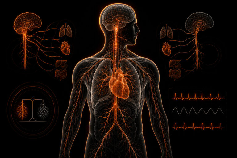
The autonomic nervous system (ANS) is the division of the peripheral nervous system responsible for involuntary physiological regulation: cardiac output, vascular tone, respiratory function, glandular secretion, and gastrointestinal motility.9 It runs continuously below conscious awareness, adjusting organ-level activity to maintain homeostasis across rest, exertion, stress, and recovery. You cannot override it directly the way you contract a skeletal muscle, but you can shift its set point indirectly through breathing patterns, exercise load, and other inputs that accumulate over time.
The ANS has two primary efferent divisions: the sympathetic nervous system, originating in the thoracolumbar spinal cord (T1-L2), and the parasympathetic nervous system, with a craniosacral origin spanning cranial nerves III, VII, IX, and X plus sacral segments S2-S4. A third division, the enteric nervous system, governs gut function semi-autonomously but sits outside the scope of the sympathetic vs parasympathetic comparison. For cardiac monitoring purposes, what matters is the interplay between the first two. Their anatomical and functional differences are the foundation for interpreting any autonomic signal you encounter in clinical or research data.
Both divisions are tonically active under resting conditions. Homeostasis emerges from their continuous interplay, not from alternating dominance. Heart rate variability (HRV) has become the most practical non-invasive index of autonomic balance in both research and monitored-care contexts, because the sinoatrial node integrates real-time input from both branches simultaneously.1 That positions HRV as a uniquely useful window into how well the two systems are calibrating against each other moment to moment.
Autonomic balance across physiological states: exercise, sleep, and stress
The sympathetic-parasympathetic ratio is not a fixed property. It shifts continuously in response to physiological demands, and understanding those shifts is what makes autonomic metrics interpretable rather than arbitrary. During vigorous aerobic exercise, sympathetic activity dominates: the heart rate climbs, vasodilation occurs in active muscle beds, vasoconstriction occurs in non-essential vascular territories, and parasympathetic tone is progressively withdrawn as intensity rises.11 At peak aerobic effort, cardiac parasympathetic input is nearly fully suppressed, which is partly why high-intensity training produces different autonomic signals than moderate-intensity exercise — parasympathetic withdrawal is near-complete in one case and partial in the other.
Recovery from exercise reverses this pattern. In the first 30-120 seconds after stopping vigorous exercise, heart rate falls rapidly: this fast component of heart rate recovery is parasympathetically mediated and reflects the speed with which vagal tone is restored after removal of the exercise stimulus. That recovery rate is itself a meaningful autonomic metric. Slowed heart rate recovery after maximal exercise — commonly defined as failure to decline by at least 12 bpm in the first minute — predicts cardiovascular mortality risk independently of resting heart rate and exercise capacity, which suggests it captures something about vagal tone restoration that other single-point measurements miss.12
Sleep produces near-complete autonomic reversal of the exercise state. During slow-wave NREM sleep, parasympathetic dominance is at its daily maximum: heart rate reaches its lowest point, HRV is highest, blood pressure dips, and the sinoatrial node receives nearly continuous vagal input that modulates each beat in the characteristic oscillating pattern captured by overnight HRV measurements. REM sleep breaks this pattern periodically, with brief surges of sympathetic activity that produce heart rate elevations and irregular HRV patterns despite the absence of physical movement. The sleep HRV profile therefore provides a dual read: parasympathetic recovery capacity during NREM and the pattern of sympathetic surges during REM, both of which carry clinical information about autonomic integrity.13
Psychological stress activates the same sympathetic pathways as physical exertion, through different mechanisms. The amygdala and hypothalamus, responding to perceived threat, drive sympathetic discharge and cortisol release, elevating heart rate, reducing HRV, and producing the cardiovascular changes that accompany anxiety, acute fear, or sustained psychological pressure.14 The physiological similarity between psychological stress activation and exercise-induced sympathetic activation is why HRV monitoring cannot distinguish between the two without contextual information. A suppressed morning HRV can reflect heavy training the previous day, a difficult night emotionally, significant life stress, illness, or poor sleep — the signal is identical, and context logging is what separates a useful interpretation from a misleading one.
Chronic sympathetic load: mechanisms and measurable consequences
When sympathetic activation is appropriate and time-limited — a stressful event followed by resolution, a hard training session followed by rest — the autonomic system returns to baseline and HRV recovers. When sympathetic activation becomes chronic, the restoration mechanism is impaired and autonomic balance shifts persistently toward sympathetic dominance. That chronic state has measurable physiological consequences beyond HRV reduction: elevated resting and nocturnal heart rate, reduced baroreflex sensitivity, attenuated heart rate recovery after exercise, and progressive reduction in parasympathetically mediated high-frequency HRV power.15
In cardiovascular terms, chronic sympathetic excess creates sustained hemodynamic load. Persistent elevation in resting heart rate — often a late consequence of reduced vagal tone rather than increased intrinsic sinus node rate — is associated with higher cardiovascular mortality risk in population studies across multiple decades of follow-up, independently of conventional risk factors.6 The relationship is not simply that sick people have higher heart rates; even in initially healthy populations, resting heart rate in the upper normal range predicts subsequent cardiovascular events at rates meaningfully higher than those observed in individuals with low resting heart rate and presumably higher vagal tone.
Chronic sympathetic dominance also interacts with inflammatory biology. Sympathetic neurotransmitters — particularly norepinephrine — modulate immune cell activity, and elevated sympathetic tone is associated with pro-inflammatory cytokine profiles in several research populations. This bidirectionality runs in both directions: inflammation itself is a reliable driver of autonomic imbalance, which is one mechanism linking chronic inflammatory conditions to elevated cardiovascular risk through autonomic pathways that sit between the inflammatory signal and the cardiac outcome.16
From a monitoring standpoint, these chronic effects are most detectable in longitudinal HRV trends rather than individual readings. A person developing chronic sympathetic load will typically show a gradual drift in baseline HRV over weeks or months, accompanied by reduced heart rate recovery after standardized exertion and reduced HRV responsiveness to recovery interventions. The signal is not a single abnormally low HRV day; it is a pattern of declining baseline that fails to respond to what should be adequate recovery. That trajectory is what continuous physiological monitoring is uniquely positioned to detect — and why platforms that provide validated, raw-accessible signal data rather than proprietary wellness scores give researchers and clinicians meaningful information about autonomic health trajectories.
Sympathetic nervous system: anatomy and physiological effects
The sympathetic branch originates from preganglionic neurons in the lateral horn of the thoracolumbar spinal cord, spanning segments T1 through L2.9 Short preganglionic neurons synapse in the paravertebral sympathetic chain ganglia or in prevertebral ganglia, and long postganglionic neurons then release norepinephrine onto alpha- and beta-adrenergic receptors at target organs. The adrenal medulla functions as a modified sympathetic ganglion, secreting epinephrine and norepinephrine directly into the bloodstream for rapid, widespread amplification of the sympathetic response when the situation demands it.
Target organ responses to sympathetic activation include: increased heart rate (positive chronotropy) and contractility (positive inotropy) via beta-1 stimulation, vasoconstriction in skin and visceral vasculature via alpha-1 receptors, vasodilation in skeletal muscle, bronchodilation, pupil dilation (mydriasis), glycogenolysis, and inhibition of digestive motility. Taken together, these effects prepare the body to respond, move, and expend energy. The system handles acute demands well. What it is not built for is chronic, sustained activation without adequate recovery between bouts.
Sustained sympathetic activation is associated with elevated allostatic load, the cumulative physiological cost of chronic stress system mobilization.4 In longitudinal cardiovascular cohort data, reduced vagal tone accompanying chronic sympathetic dominance is independently associated with adverse cardiovascular outcomes.3 The distinction between acute sympathetic response (adaptive) and chronic sympathetic load (costly) is one of the most practically important points to carry forward from this physiology, and it directly shapes how clinicians and researchers should interpret sustained low-HRV patterns over time.
Parasympathetic nervous system: anatomy and physiological effects
The parasympathetic nervous system has a craniosacral distribution that looks quite different from the sympathetic system’s thoracolumbar origin. Cranial outflow travels via four cranial nerves: CN III (pupil constriction and lens accommodation), CN VII (lacrimal and salivary glands), CN IX (parotid gland), and CN X, the vagus nerve. Sacral outflow via S2-S4 innervates pelvic organs.9 The vagus nerve carries roughly 75% of all parasympathetic fibers and is the dominant parasympathetic pathway to the heart, lungs, and abdominal viscera. Its reach is broad, and its influence on cardiac function is direct and measurable in real time.
Postganglionic neurons throughout this system release acetylcholine onto muscarinic receptors. At the sinoatrial node, this produces negative chronotropy (reduced heart rate) and slowed AV conduction. Other target effects include enhanced gastrointestinal motility, increased glandular secretion, mild bronchoconstriction, and pupil constriction (miosis). The net picture is a body conserving resources, restoring tissue, and returning to a calibrated resting state after demand. That restorative capacity is what makes parasympathetic function so central to recovery science and longitudinal health monitoring.
Vagal tone describes the sustained level of acetylcholine release at the sinoatrial node and is directly reflected in high-frequency HRV power and RMSSD values.2 Higher vagal tone generally indicates a heart that responds more flexibly to moment-to-moment physiological input. For a closer look at what those measurements actually reflect and where their interpretive limits lie, see vagal tone meaning: signal, noise, and measurement limits. That said, one popular theoretical framework warrants a note of caution here: polyvagal theory has proposed a hierarchical model of vagal regulation with evolutionary structure, but this framework remains actively debated in peer-reviewed literature and should not be treated as settled autonomic physiology.6 For a full review of the evidence base and where the data actually stands, see polyvagal theory: evidence, accuracy, and clinical use.
Sympathetic vs parasympathetic: key differences at a glance
The sympathetic vs parasympathetic systems differ across every major organizational axis: anatomical origin, neurotransmitter profile, receptor types, target organ responses, and cardiovascular direction.9 The table below captures the core contrasts. One important caveat applies before reading it: neither system works as a simple on-off switch. Co-activation occurs across many physiological states, and HRV reflects the continuous integrated output of both branches rather than a clean ratio between them.1 The table is a map of functional differences, not a description of alternating dominance.
| Feature | Sympathetic Branch | Parasympathetic Branch |
|---|---|---|
| Spinal origin | Thoracolumbar (T1-L2) | Craniosacral (CN X; S2-S4) |
| Postganglionic neurotransmitter | Norepinephrine | Acetylcholine |
| Heart receptor type | Beta-1 adrenergic | Muscarinic (M2) |
| Effect on heart rate | Increases (positive chronotropy) | Decreases (negative chronotropy) |
| Primary HRV proxy | Low HF power; reduced RMSSD | High HF power; elevated RMSSD |
| Metabolic role | Glycogenolysis; lipolysis | Promotes anabolic recovery |
| Response onset at heart | Seconds | Seconds to minutes |
How HRV reflects the balance between sympathetic and parasympathetic activity
Heart rate variability quantifies the beat-to-beat variation in R-R intervals (ECG) or inter-beat intervals (PPG-derived).1 Because the sinoatrial node integrates simultaneous sympathetic vs parasympathetic input in real time, HRV analysis can partially decompose their respective contributions. RMSSD (root mean square of successive differences) is the primary time-domain index of vagal (parasympathetic) modulation. SDNN, by contrast, captures total autonomic variability and reflects input from both branches.2 That difference matters more than most introductory HRV content acknowledges. For a signal-level explanation of what the R-R interval actually measures versus what PPG-derived pulse intervals capture, see how r-r interval vs pulse interval is computed at the signal level.
Frequency-domain decomposition separates HRV into spectral bands. The high-frequency (HF) band (0.15-0.40 Hz) corresponds to respiratory sinus arrhythmia and is considered predominantly parasympathetic. The low-frequency (LF) band (0.04-0.15 Hz) has mixed neural origins. The LF/HF ratio is frequently cited as a sympathovagal balance metric, but this interpretation is actively contested: LF power reflects baroreflex dynamics, respiratory coupling, and other non-sympathetic inputs, and the ratio does not cleanly isolate sympathetic efferent activity.6 Treating LF/HF as a direct readout of sympathetic tone is one of the most common and consequential misreads in applied autonomic research. The evidence does not support it.
A 2024 meta-analysis of vagally mediated HRV confirmed that RMSSD and HF power are the most reliable autonomic markers across field recording conditions.5 Continuous PPG platforms that derive HRV from raw optical waveforms make longitudinal ANS monitoring across days to weeks possible in a way that episodic clinical recordings simply cannot match. Learn how Sensor Bio’s Pulse Engine extracts HRV metrics from continuous PPG waveforms.
Clinical relevance: what reduced vagal tone and chronic sympathetic load signal
Low HRV, as a marker of reduced parasympathetic modulation, is independently associated with increased all-cause and cardiovascular mortality in longitudinal cohort studies, even after adjustment for conventional risk factors.7 This association is not isolated to a single population or study design. It appears consistently across clinical groups: reduced vagal tone is documented in post-myocardial infarction patients, individuals with heart failure, those with type 2 diabetes, and chronic pain populations compared to age-matched controls.3 That consistency matters, because it shifts the finding from one interesting cohort result toward a broader, reproducible physiological pattern.
Sustained sympathetic nervous system activation is associated with elevated allostatic load, the cumulative physiological cost of chronic stress response mobilization.4 Several modifiable factors are associated with improved vagal tone in group studies: regular aerobic exercise, slow-paced breathing protocols, and adequate sleep duration. Heart rate recovery speed after submaximal exertion serves as a validated field index of post-exercise vagal reactivation.10 You can also explore related physiology in parasympathetic saturation explained: what the research shows, which goes further into the boundaries of parasympathetic measurement and what happens at the edges of vagal capacity.
These are population-level statistical associations from epidemiological cohorts, not individual diagnostic thresholds. Low HRV reflects a physiological pattern associated with elevated risk at the group level and does not constitute a stand-alone diagnostic criterion for any specific condition. For a deeper review: low HRV: clinical significance and what the evidence shows and how HRV and resting heart rate measure different autonomic dimensions.
Measuring autonomic activity: ECG, PPG, and signal considerations
The reference standard for assessing the sympathetic vs parasympathetic balance via cardiac signals is ECG-derived HRV, using R-R intervals sampled at a minimum of 250 Hz (1000 Hz preferred for high-precision research).1 ECG directly measures cardiac electrical activity, providing millisecond-level R-R interval precision under controlled conditions. Standard 5-minute resting recordings in a controlled posture are the minimum for frequency-domain analysis. Twenty-four-hour Holter recordings go considerably further, enabling full circadian autonomic profiling that a brief resting assessment cannot produce.
PPG-derived HRV uses optical pulse detection to estimate inter-beat intervals (IBI) from pulse wave peaks. PPG has been validated against ECG for key HRV metrics including RMSSD and HF power, with documented accuracy limits under conditions of motion artifact, low perfusion pressure, skin tone variability, and inconsistent sensor contact.8 Ultra-short recordings of one to five minutes provide valid RMSSD estimates in field settings. Full frequency-domain decomposition requires at least five minutes under standardized conditions.5 Knowing where PPG is reliable and where it breaks down is as important as knowing what it measures, particularly when interpreting autonomic data collected outside a controlled clinical setting.
Other ANS assessment approaches include electrodermal activity (reflecting sympathetic sudomotor function), blood pressure variability, baroreflex sensitivity testing, and pupillometry. Each captures a distinct aspect of autonomic control and should not be treated as interchangeable with HRV-based metrics. The choice of method should follow the physiological question, not convenience. For context on the signal quality factors that affect PPG accuracy in field conditions: PPG signal quality and the factors that affect HRV accuracy.
Limits and pitfalls when interpreting autonomic metrics
Several recurring misinterpretations arise when applying sympathetic vs parasympathetic concepts to HRV data or other autonomic measures. These are not edge-case errors. They appear regularly in applied research, clinical summaries, and monitoring platform documentation alike. Clinicians and researchers working with autonomic biomarkers should account for each of the following when drawing conclusions about autonomic state from a single measurement session or a short recording window:
- LF/HF ratio as a direct sympathovagal balance index: LF power has multiple neural and mechanical origins, not sympathetic efferents alone. Using LF/HF as a direct sympathetic activity proxy is not well-supported by current evidence and has been explicitly critiqued in peer-reviewed autonomic physiology literature.6
- PPG-derived HRV as equivalent to ECG-derived HRV: The two modalities are not interchangeable without qualification. Validation studies show reasonable agreement for RMSSD and HF power under controlled conditions, but diverge substantially with motion artifact, peripheral vasoconstriction, high-frequency noise, or inadequate sampling rates.8
- Population reference ranges applied to individuals: HRV normative data from published standards describes population-level distributions, not individual diagnostic cutoffs.2 A single low RMSSD reading does not indicate autonomic dysfunction. Longitudinal within-individual tracking across comparable conditions carries substantially more interpretive weight than single-point comparisons to population norms.
- Polyvagal theory as established consensus physiology: The polyvagal framework proposes a hierarchical vagal model with evolutionary structure. It remains actively contested in peer-reviewed literature and should be referenced as a theoretical model rather than empirical fact.6
FAQ
What is the main difference between the sympathetic and parasympathetic nervous system?
In the sympathetic vs parasympathetic pairing, the sympathetic nervous system mobilizes the body for action: it increases heart rate, elevates blood pressure, redirects blood flow to skeletal muscle, dilates bronchioles, and suppresses digestive activity. The parasympathetic nervous system drives recovery and restoration: it decreases heart rate, promotes gastrointestinal motility, stimulates glandular secretion, and supports tissue repair. Both systems are tonically active at rest, and homeostasis depends on their continuous dynamic interplay rather than one simply overriding the other. The vagus nerve (cranial nerve X) carries the majority of parasympathetic output to the heart, lungs, and abdominal viscera.9 Neither system is meant to be permanently dominant. Regulatory flexibility, meaning the capacity to shift appropriately between states and return to baseline, is the actual measure of autonomic health in both clinical and research contexts.
Can you have too much sympathetic or parasympathetic activity?
Yes, imbalance in either direction carries measurable physiological consequences. Chronic sympathetic dominance, often indexed by persistently low HRV, is associated with elevated cardiovascular risk, impaired immune regulation, and metabolic stress in longitudinal cohort studies.7 Excessive parasympathetic tone (vagotonia) can produce bradycardia, hypotension, or vasovagal syncope in susceptible individuals. The clinical goal is not maximum activation of either branch but regulatory flexibility: the capacity to shift appropriately between states and return to baseline. Serial HRV monitoring is one practical way to quantify that flexibility over time, rather than relying on a single-point snapshot that may not reflect the person’s typical autonomic range.
What does HRV measure in relation to sympathetic vs parasympathetic balance?
HRV captures beat-to-beat variation in the sinoatrial node’s firing interval, which integrates real-time input from both sympathetic and parasympathetic pathways simultaneously. RMSSD and HF power (0.15-0.40 Hz) are the most direct and reliable vagal (parasympathetic) indices in field recording conditions.5 SDNN reflects total autonomic variability from both branches. The LF/HF ratio is frequently cited as a sympathovagal balance metric, but this interpretation is contested: LF power has mixed neural origins including baroreflex dynamics and respiratory coupling, and it does not cleanly isolate sympathetic efferent activity.6 Treating RMSSD as your primary vagal index and SDNN as your total variability measure is the more defensible approach in both research and clinical monitoring contexts.
Does exercise shift the balance between sympathetic and parasympathetic activity?
Yes, with direction depending on phase. During aerobic exertion, sympathetic drive increases and parasympathetic withdrawal is rapid. During recovery, the rate of vagal reactivation becomes visible as the heart rate recovery slope: faster recovery reflects more efficient parasympathetic reinstatement and is a validated measure of cardiovascular fitness.10 Regular aerobic training is consistently associated with elevated resting vagal tone in group studies, reflected as higher resting RMSSD and HF power. This adaptation is generally more pronounced with endurance training than with resistance work alone. That said, these are population-level patterns. Individual responses depend on training volume, intensity, and baseline autonomic function, which is why longitudinal tracking across consistent recording conditions carries more interpretive value than comparing a single session to population norms.
How does PPG compare to ECG for measuring autonomic activity?
ECG directly measures cardiac electrical activity and is the reference standard for R-R interval-based HRV analysis, providing millisecond-level precision in controlled conditions.1 PPG detects peripheral pulse waveforms optically and derives inter-beat intervals from peak detection. PPG-based vagal HRV indices (RMSSD, HF power) have been validated against ECG under controlled conditions, but the two modalities are not interchangeable without qualification.8 Motion artifact, peripheral perfusion variability, skin tone effects, and sampling rate differences all affect inter-beat interval accuracy in real-world field use. PPG’s primary advantage is enabling continuous longitudinal monitoring across days and weeks, capturing autonomic patterns that a brief clinical recording cannot see. For single-session high-precision autonomic assessment, ECG remains the reference method.
Is low HRV the same as sympathetic dominance?
Not precisely. Low HRV primarily indicates reduced parasympathetic (vagal) modulation of the sinoatrial node rather than a direct measure of elevated sympathetic tone.2 Reduced vagal modulation and relatively increased sympathetic influence often co-occur, but HRV metrics do not provide a clean isolated measure of sympathetic efferent activity. RMSSD and HF power are vagal indices: they reflect how much the parasympathetic branch is modulating the heart’s pacemaker cycle-to-cycle. Low RMSSD is associated with increased cardiovascular risk at the population level but is not a diagnostic criterion for sympathetic dominance or any specific clinical condition without broader evaluation.
For research groups, clinical programs, and occupational health teams evaluating continuous autonomic nervous system monitoring platforms, learn how Sensor Bio’s research-grade PPG platform supports longitudinal HRV data collection.
References
References
- Task Force of the European Society of Cardiology and the North American Society of Pacing and Electrophysiology. Heart Rate Variability: Standards of Measurement, Physiological Interpretation and Clinical Use. Circulation. 1996;93(5):1043-1065. DOI: 10.1161/01. CIR.93.5.1043
- Shaffer F, Ginsberg JP. An Overview of Heart Rate Variability Metrics and Norms. Frontiers in Public Health. 2017;5:258. DOI: 10.3389/fpubh.2017.00258
- Thayer JF, Lane RD. The Role of Vagal Function in the Risk for Cardiovascular Disease and Mortality. Biological Psychology. 2007;74(2):224-242. DOI: 10.1016/j.biopsycho.2005.11.013
- McEwen BS. Protective and Damaging Effects of Stress Mediators. New England Journal of Medicine. 1998;338(3):171-179. DOI: 10.1056/NEJM199801153380307
- Laborde S, Allen MS, Borges U, et al. Vagally Mediated Heart Rate Variability and Indexed Measures of Cognitive and Emotional Processes: A Meta-analysis. Clinical Physiology and Functional Imaging. 2024. DOI: 10.1111/cpf.12862
- Grossman P. A Quarter Century of Polyvagal Theory: Do Recent Data Provide Support? Biological Psychology. 2023;174:108389. DOI: 10.1016/j.biopsycho.2022.108389
- Dekker JM, Crow RS, Folsom AR, et al. Low Heart Rate Variability in a 2-Minute Rhythm Strip Predicts Risk of Coronary Heart Disease and Mortality: The ARIC Study. Circulation. 2000;102(11):1239-1244. DOI: 10.1161/01. CIR.102.11.1239
- Allen J. Photoplethysmography and Its Application in Clinical Physiological Measurement. Physiological Measurement. 2007;28(3):R1-R39. DOI: 10.1088/0967-3334/28/3/R01
- Waxenbaum JA, Reddy V, Varacallo M. Anatomy, Autonomic Nervous System. In: StatPearls. Treasure Island (FL): StatPearls Publishing; 2023. PMID: 30860696. Available at: https://www.ncbi.nlm.nih.gov/books/NBK539845/
- Shetler K, Marcus R, Froelicher VF, et al. Heart Rate Recovery: Validation and Methodologic Issues. Journal of the American College of Cardiology. 2001;38(7):1980-1987. DOI: 10.1016/S0735-1097(01)01652-7