Updated: May 15, 2026
low hrv symptoms are not diagnosed by a wearable alone; the more useful pattern is persistent low HRV paired with fatigue, poor sleep, high resting heart rate, illness, overtraining, or new symptoms.
low hrv symptoms verification checklist
- Compare HRV to the person’s own baseline rather than a population average.
- Check sleep duration, alcohol, medication changes, infection, training load, and mental stress before interpreting the signal.
- Escalate new chest pain, fainting, severe shortness of breath, or sustained palpitations to medical evaluation.
Related Sensor Bio reading: how to improve HRV, vagal tone meaning, and HRV and inflammation.
Helpful references include a Frontiers review of HRV physiology, PubMed research on HRV and fatigue symptoms, and Mayo Clinic guidance on heart-rate context.
Low heart rate variability symptoms are not sensations you feel in the moment. They are physiological patterns, including fatigue, poor sleep recovery, and reduced autonomic flexibility, that research associates with the chronic ANS dysregulation low HRV reflects rather than causes.1 That distinction is worth sitting with before you interpret your next reading, because the difference between HRV as a cause and HRV as a reflection changes everything about what you should do with the data.
When you notice a depressed HRV reading, the instinct is to search for a matching checklist of symptoms. That framing inverts the relationship. What the scientific literature actually documents is a pattern: conditions that chronically suppress parasympathetic nervous system tone produce observable downstream consequences in how your body sleeps, recovers from physical effort, and adapts to psychological pressure. The low HRV reading and the fatigue you notice after a difficult week are both outputs of the same ANS state, not cause and effect. Understanding what HRV actually measures, and what it cannot tell you, is the foundation for reading these signals accurately rather than catastrophizing a single data point.
This article covers the physiological mechanism behind HRV, the research associations between suppressed HRV and common symptom experiences, how wearable optical sensors measure HRV and where they fall short, and what a persistently low HRV trend should and should not prompt you to do. The goal is not to reassure or alarm. It is to give you the evidence-grounded framework to read your own data more clearly.
What Is HRV and Why Does a Low Value Matter?
Heart rate variability is the millisecond-level fluctuation in timing between consecutive heartbeats, measured from the R-R interval on an ECG or the equivalent pulse-to-pulse interval on an optical sensor. Those fluctuations are not random noise. They arise from the continuous push-pull between the two branches of the autonomic nervous system: the parasympathetic branch, which drives beat-to-beat variability upward, and the sympathetic branch, which suppresses it during stress mobilization. If you want a direct comparison between HRV and the simpler metric of resting heart rate, and what each one actually tells you about cardiovascular and autonomic health, the breakdown in HRV vs. resting heart rate covers the clinical distinction in full.
Standard HRV metrics divide into time-domain and frequency-domain measures. RMSSD, the root mean square of successive R-R interval differences, captures short-term parasympathetic tone and is the most widely used metric in wearable monitoring. SDNN, the standard deviation of all normal-to-normal intervals, reflects broader ANS activity over longer recordings. LF and HF spectral band power separate sympathetic and parasympathetic contributions, respectively.1 Each metric answers a slightly different question about your autonomic state, and which one matters most depends on what you are monitoring and over what time window. A single overnight RMSSD reading and a 24-hour Holter-derived SDNN are measuring related but distinct aspects of autonomic function.
A chronically low HRV value signals reduced autonomic flexibility: the ANS is not adapting dynamically to internal and third-party demands. This is not a direct indicator of any specific disease. It is a well-validated physiological marker associated with states that correlate with what people describe as low heart rate variability symptoms, including persistent fatigue, reduced stress tolerance, and slow recovery from both physical and psychological effort. The signal is real. The causal direction, however, is more nuanced than most popular health content suggests, and the distinction shapes both how you interpret your data and how you respond to it.
How lifestyle factors and training load interact with HRV
Low HRV readings are not always signs of something wrong. They can also be the expected output of specific choices — and recognizing that distinction is essential for anyone using HRV as a feedback tool. Acute training stress is among the most reliable ways to temporarily suppress HRV. A hard endurance session or heavy resistance training block commonly reduces next-morning HRV by 10-25% from an individual’s established baseline, with the degree of suppression correlating loosely with training volume and intensity.7 That short-term suppression is not a symptom of dysfunction; it is the expected physiological response to a significant metabolic load, reflecting the sympathetic activation and parasympathetic withdrawal that accompany muscle repair and adaptation.
The problem arises when that suppression fails to resolve. In athletes accumulating a planned training block — called functional overreaching — HRV typically recovers over 24-72 hours following rest. In non-functional overreaching and early overtraining syndrome, HRV remains persistently low for weeks, often accompanied by sleep disruption, reduced motivation, and performance plateau or decline.8 Those persistent low HRV readings are not measurement artifacts; they reflect genuine autonomic dysregulation from accumulated physiological stress that has exceeded the body’s capacity for adaptive recovery. The trajectory — whether HRV is recovering across days or staying suppressed — matters more than any single low reading.
Sleep deprivation produces overlapping effects. Even a single night of significantly reduced sleep (less than 5-6 hours in a habitually 7-8 hour sleeper) is enough to suppress next-day HRV measurably, primarily through reduction in parasympathetic tone and elevation of cortisol and sympathetic activity.9 This creates a common interpretation problem in self-monitoring: the low-HRV morning that follows poor sleep can look identical to the low-HRV morning that follows hard training. Context tracking — noting sleep hours, training load, and perceived stress alongside HRV readings — is what separates one from the other, not the number itself.
Alcohol is another behavioral factor that reliably suppresses HRV in ways that are detectable even from modest intake the night before. Research using controlled alcohol administration shows dose-dependent HRV reduction that persists through the following morning, with next-day RMSSD values measurably lower even after mild to moderate drinking.10 The mechanism involves both direct cardiac autonomic suppression and the fragmented, REM-suppressing sleep that follows alcohol consumption. From a monitoring standpoint, this means that HRV readings collected without accounting for alcohol intake can be systematically biased downward in ways that obscure true physiological trends.
Taken together, these behavioral influences on HRV point toward the same core requirement: interpretation in context. A single low HRV reading is noise. A pattern of low HRV that co-occurs with identifiable stressors and resolves with recovery is signal. A persistent low HRV trend that resists recovery despite reduced training and adequate sleep is a different kind of signal — one that warrants clinical evaluation. That graded interpretation framework is what distinguishes useful HRV monitoring from pattern-matching anxiety over individual numbers. For more on what evidence-based interventions can shift HRV upward over time, how to raise heart rate variability covers the published literature in detail.
Measurement differences that can produce artificially low HRV readings
Not every low HRV value reflects low HRV. Signal quality, recording conditions, and device-specific processing choices can all produce suppressed HRV outputs that have nothing to do with the user’s actual cardiac autonomic state. Understanding the main sources of artifact is especially relevant for PPG-based wrist monitors, where signal quality is more variable than ECG under real-world conditions.
Motion artifact is the primary culprit during active or restless sleep. PPG relies on stable optical contact with the skin to accurately detect the peak of each pulse wave, and even minor wrist movement can shift the waveform in ways that displace detected peaks and distort beat-to-beat interval timing. HRV calculated from artifact-contaminated intervals will appear lower than actual HRV, because erroneously long or short intervals inflate interval variability in ways that algorithms may not fully correct.5
Poor sensor contact, cold ambient temperature (which causes peripheral vasoconstriction and reduces PPG signal amplitude), arrhythmias, and ectopic beats all create similar problems. An ectopic beat inserts an abnormally short or long R-R interval that dramatically inflates variability metrics if included, or produces an anomalously low RMSSD if the algorithm clips it incorrectly. Many consumer devices attempt to filter ectopics, but filter implementations vary widely and are rarely disclosed. The same underlying autonomic physiology can produce notably different HRV values on different devices or even different software versions of the same device.
This is why, for any low HRV reading that seems inconsistent with how someone feels or what they have been doing, the first question is whether the measurement is technically clean. Artifacts in a 5-minute resting ECG recording are visible to a trained reader; artifacts in a wearable PPG-derived HRV value are often invisible to the user. Platforms that provide raw waveform access alongside derived HRV metrics allow researchers and clinicians to inspect signal quality independently, which is a meaningful differentiator for any application where measurement accuracy matters.6
Physiological and Clinical Associations: What Research Links to Low HRV
Several physiological domains consistently correlate with chronically low HRV in peer-reviewed literature. These are epidemiological associations grounded in ANS dysregulation, not established causal chains. Low HRV reflects an underlying state; it does not itself produce the experiences described below. Knowing this difference changes how you read your data and what you do with it, because chasing the HRV number without addressing the underlying condition is treating the indicator rather than the problem.
Chronic stress and allostatic load. Sustained psychological or physiological stress elevates cortisol, which suppresses vagal tone and reduces parasympathetic-driven HRV. Research on populations experiencing chronic occupational or psychological stress shows consistent RMSSD reduction relative to lower-stress controls.2 The mechanism is direct: when the stress response is chronically activated, the ANS privileges sympathetic drive over parasympathetic recovery, and RMSSD reflects that shift in real time. Conditions associated with chronically low HRV in this domain include persistent fatigue, brain fog, and blunted emotional regulation, all of which are downstream effects of the same autonomic suppression, not independent consequences of seeing a low number on your screen.
Cardiovascular risk. In landmark prospective data, Kleiger et al. (1987, American Journal of Cardiology) demonstrated that post-myocardial infarction patients with lower SDNN faced significantly elevated all-cause mortality, establishing SDNN as an independent predictor of adverse cardiovascular outcomes.3 This remains one of the most cited findings linking low HRV to clinical risk. It is also worth noting that this association emerged from 24-hour Holter recordings in a post-MI clinical population, not from single-morning wrist-sensor readings in otherwise healthy people. Context and measurement protocol matter enormously when you try to translate population-level risk data into personal clinical decisions.
Post-viral and long-COVID dysautonomia. A 2024 systematic review in the Journal of Electrocardiology (Ferreira et al.) documented persistent RMSSD depression in long-COVID cohorts compared to matched controls, with autonomic dysfunction proposed as a central mechanism linking post-viral fatigue and poor recovery capacity to suppressed parasympathetic activity.4 Symptoms documented in these cohorts included exercise intolerance, orthostatic sensitivity, and disrupted sleep, including patterns consistent with low heart rate during sleep and unrefreshing rest, reflecting the same autonomic disruption that suppresses daytime HRV. The breadth of this evidence now supports viewing dysautonomia as a genuine component of long-COVID pathophysiology, not a fringe hypothesis.
Athletic overtraining. Sustained parasympathetic withdrawal, visible as a declining multi-day RMSSD trend, is a well-documented signature of insufficient recovery in endurance athletes. Subjective correlates include reduced motivation, elevated perceived effort at submaximal workloads, and mood disturbances. Some athletes also experience disrupted nocturnal autonomic regulation, with periods of elevated heart rate during sleep reflecting the same sympathetic overdrive that suppresses daytime HRV. The multi-day RMSSD trend provides an earlier and more objective signal of this overreached state than subjective rating scales alone.
Psychiatric populations. Depression and anxiety disorders are associated with reduced vagal tone and lower RMSSD values in meta-analytic evidence.2 The ANS dysregulation in these conditions produces measurable suppression of parasympathetic activity, which manifests in the HRV signal. The magnitude of HRV reduction appears to scale with symptom severity in several studies, which has led researchers to propose HRV normalization as a potential biomarker for treatment response. That application remains in active investigation, but the foundational link between psychiatric symptom burden and HRV suppression is well-supported across multiple independent datasets.
Metabolic dysfunction and cardiovascular risk: key cohort findings
Two major longitudinal cohorts established some of the clearest epidemiological links between low HRV and clinical risk. The Framingham Heart Study documented that hyperglycemia is associated with reduced HRV, demonstrating how metabolic dysfunction directly impairs autonomic regulation. This finding underscores why HRV monitoring may carry additional relevance in pre-diabetic and diabetic populations beyond its general wellness application.
The ARIC (Atherosclerosis Risk in Communities) study found that reduced cardiac autonomic function could predict the manifestation of hypertension up to three years later in initially normotensive individuals. This prospective finding positions low HRV as a potential early indicator of developing cardiovascular risk, not merely a correlate of existing disease. Type 2 diabetes is similarly associated with reduced HRV, particularly when complications such as peripheral neuropathy are present, reflecting progressive autonomic fiber damage.
These population associations support interpreting a chronically low HRV trend as a signal warranting broader clinical assessment, not as a standalone diagnostic finding.
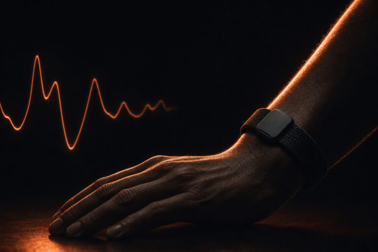
How HRV Is Measured: ECG, PPG, and the Wrist-Sensor Accuracy Question
The clinical reference standard for HRV measurement is the electrocardiogram (ECG). ECG electrodes detect the precise electrical R-wave peak with millisecond-level accuracy, and R-R intervals computed from these peaks form the basis of all validated HRV metrics defined in the 1996 Task Force standard.1 When you see HRV reference ranges quoted in clinical literature, they were almost always derived from ECG recordings in controlled laboratory or Holter conditions, not from wrist-worn optical sensors during daily life. That provenance matters when you try to translate population-level clinical evidence into personal wearable data.
Wrist-worn optical sensors use photoplethysmography (PPG): a light source shines into the skin, and a photodetector measures reflected light, capturing changes in blood volume with each heartbeat. The timing between successive pulse peaks, called the pulse-to-pulse interval (PPI), approximates the ECG R-R interval. PPI-derived RMSSD correlates with ECG-derived RMSSD under controlled, low-motion conditions. To understand how the autonomic nervous system and wearable HRV measurement interact at a deeper physiological level, the wearable HRV and autonomic nervous system overview covers the mechanistic layers behind what these sensors actually detect.
Li et al. (2023, iScience) evaluated wearable PPG-derived HRV accuracy across diverse populations and found moderate-to-strong agreement with ECG reference in resting and low-motion states.5 Costantini et al. (2023, Sensors) confirmed that wrist-worn optical sensors can yield valid RMSSD estimates when sampling rate, sensor-to-skin contact quality, and motion exposure are adequately controlled.6 Accuracy degrades under high motion, low skin perfusion, and certain cardiac arrhythmias. For arrhythmia-prone populations especially, PPG-derived HRV should be interpreted with full awareness of this limitation. Skin tone and perfusion also affect optical signal quality in ways that vary by sensor design and wavelength; the evidence on how different skin tones influence PPG measurement accuracy is examined in the PPG skin tone validation research summary.
That said, accuracy limitations do not make wrist-based PPG useless. Researchers and clinical teams evaluating remote therapeutic monitoring (RTM) workflows are increasingly exploring PPG-based optical sensors for continuous, longitudinal HRV data collection outside the clinic, where the convenience of passive wrist-based monitoring and high patient compliance offset the modest accuracy reduction compared to ECG for trend-level analysis. The key is understanding what you are measuring, where the limits are, and what decisions that data can and cannot responsibly support.
What a Single Low Reading Means (and Does Not Mean)
Day-to-day HRV is inherently noisy. Within-person variability of 20 to 40 percent across a single week is normal, driven by factors including alcohol consumption, acute illness, sleep debt, ambient temperature, and, in people who menstruate, cycle phase.1 A single depressed reading is rarely meaningful on its own. This is one of the most important things to internalize early if you are new to HRV monitoring: the metric is designed for longitudinal interpretation, not read like a blood pressure cuff on any given morning.
The actionable signal lies in trends, not snapshots. A persistent downward shift in your rolling 7-to-14-day RMSSD baseline, relative to your own established historical norm, carries far more informational weight than any individual data point or any comparison to population reference values. This is precisely why both research protocols and clinical monitoring programs consistently emphasize individual baseline tracking over absolute thresholds. If you see one low reading after a late night or a stressful day, you are observing exactly what HRV is supposed to detect: a transient shift in autonomic state. That is the metric working correctly, not a warning sign that demands intervention.
The second interpretive error to avoid is comparing your HRV to published third-party norms. Population-level HRV reference ranges vary substantially by age, sex, fitness level, and measurement protocol.1 A RMSSD that falls below average for a 30-year-old in good cardiovascular health may be entirely typical for a healthy 60-year-old. The most actionable signal is a meaningful, sustained deviation from your own rolling average, correlated with subjective changes in energy, sleep quality, or performance. Chasing a published population number is not only unhelpful; it can lead you to treat a completely normal physiological state as a problem requiring intervention, which is a misuse of the data.
Which Populations Show Low HRV Most Reliably in Research
Research has documented consistent HRV suppression across several specific populations. Understanding which groups are best studied helps contextualize what low heart rate variability symptoms look like at the population level, and it also clarifies the difference between HRV patterns that are normal for a given life stage and those that reflect genuine pathological disruption worth investigating clinically.
Older adults. HRV declines with age even in healthy, fit individuals. SDNN and RMSSD reductions across the lifespan are normative physiological phenomena, not inherently pathological signals when they occur in isolation.1 Aging-related HRV decline reflects reduced autonomic range rather than disease. An older adult with chronically low absolute RMSSD is not necessarily more at risk than a younger person with higher absolute values; what matters clinically is the trajectory relative to that individual’s own history and the concurrent clinical picture, not the comparison to a population percentile from a published reference table.
Post-myocardial infarction populations. Post-MI populations show the strongest, most consistently documented link between low SDNN and adverse cardiovascular outcomes in longitudinal data. SDNN values below 50 ms in 24-hour Holter recordings have been associated with significantly elevated mortality risk in this group.3 This association was established in controlled clinical settings using gold-standard ECG measurement over full 24-hour periods, and it should not be casually extrapolated to wrist-sensor readings from otherwise healthy individuals. The clinical significance of low HRV is highly context-dependent, and the post-MI population represents one of the best-evidenced high-risk contexts.
Long-COVID and dysautonomia. Ferreira et al. (2024, Journal of Electrocardiology) synthesized data from multiple long-COVID cohort studies and found significantly lower RMSSD values compared to controls, consistent with persistent dysautonomia as a post-viral sequela.4 Reported symptom correlates in these cohorts include fatigue, post-exertional malaise, and impaired orthostatic tolerance. The consistency of this finding across independent cohort studies strengthens the case for autonomic dysfunction as a mechanistically meaningful component of long-COVID, not simply a symptom cluster of unclear etiology.
Depression and anxiety populations. Meta-analytic evidence links depression and anxiety disorders to reduced vagal tone and lower resting RMSSD.2 The magnitude of HRV reduction scales with symptom severity in several studies, and HRV normalization has been proposed as a potential biomarker for treatment response. If your HRV trend worsens in parallel with declining mood or increasing anxiety, the two signals are most likely reflecting the same underlying autonomic dysregulation rather than one causing the other. That framing matters for how you prioritize your response.
Autonomic neuropathy populations. Individuals with autonomic neuropathy secondary to chronic systemic disease show among the most pronounced HRV reductions in the research literature, reflecting direct damage to autonomic regulatory pathways rather than functional suppression alone. In these populations, HRV monitoring serves a different clinical purpose: tracking disease progression and treatment response rather than optimizing recovery or flagging transient stressors. The distinction between functional autonomic suppression and structural autonomic damage is one of the more important interpretive lines to hold when working with HRV data in clinical populations.
When to Seek Professional Evaluation
HRV data from wearable monitoring is best understood as a prompt for clinical conversation, not as a diagnostic instrument. A persistent downward trend in your HRV baseline, sustained over two or more weeks despite adequate rest, is worth discussing with a clinician, particularly if accompanied by unexplained fatigue, reduced exercise capacity, or a known history of cardiovascular, post-viral, or autonomic conditions. For a thorough look at what the research shows about the causes, cardiovascular risk associations, and evidence-based approaches to improving low HRV, the low heart rate variability: causes, risks, and how to improve HRV overview covers the full evidence base in depth.
Within clinical practice, Remote Therapeutic Monitoring (RTM) under CPT codes 98975 through 98981 provides a structured framework for licensed practitioners to review and act on physiological data collected outside the clinic. RTM is distinct from Remote Patient Monitoring (RPM, codes 99453 through 99458) in both scope and documentation requirements; the two should not be conflated. Any RTM engagement requires licensed provider oversight of data interpretation and clinical decision-making. Clinics and health systems evaluating whether to incorporate wearable biosignal data into remote care programs can find a structured vendor evaluation framework at remote patient monitoring companies: vendor evaluation framework for clinics.
Sensor Bio’s optical wearable platform is not FDA-cleared as a diagnostic device. Consumer self-monitoring of HRV provides personal trend context and can support informed conversations with providers, but it does not constitute clinical assessment. If you experience chest pain, syncope, severe shortness of breath, or significant new changes in cardiac rhythm, seek emergency evaluation immediately, regardless of your HRV readings.
Evidence Summary: Key Research on HRV and Its Clinical Associations
| Claim | HRV Metric | Direction | Citation |
|---|---|---|---|
| HRV is a validated ANS balance marker; RMSSD and SDNN are established standards | RMSSD, SDNN, LF/HF | Established standard | Task Force ESC/NASPE 19961 |
| Depression and anxiety are associated with reduced vagal tone and lower RMSSD | RMSSD | Reduced in affected populations | Kemp et al. 20122 |
| Lower SDNN after myocardial infarction is associated with elevated all-cause mortality | SDNN | Inverse relationship with survival | Kleiger et al. 19873 |
| Long-COVID cohorts show persistent RMSSD depression compared to controls | RMSSD | Reduced vs. matched controls | Ferreira et al. 20244 |
| Wrist PPG-derived HRV correlates with ECG-derived RMSSD in controlled, low-motion conditions | PPI-derived RMSSD | Moderate-to-strong agreement | Li et al. 2023; Costantini et al. 202356 |
FAQ
What are the most common symptoms associated with low heart rate variability symptoms?
Low HRV is a marker of reduced autonomic nervous system flexibility, not a direct cause of specific sensations. Conditions that research consistently links to chronically suppressed HRV, including high allostatic stress load, post-viral autonomic dysfunction, and cardiovascular disease, are associated with fatigue, poor sleep quality, brain fog, and slow physical recovery.24 The underlying condition produces the symptom experience; the low HRV reading reflects the same physiological disruption from a different angle. Treating low HRV as the cause rather than the indicator leads to misinterpretation of what the signal is telling you, and often to misplaced intervention.
Is low HRV dangerous?
A single low HRV reading is rarely meaningful in isolation. Sustained low SDNN over weeks has been associated with elevated cardiovascular risk in large prospective cohort studies, particularly in post-MI populations.3 Persistently low RMSSD has also been documented in long-COVID dysautonomia cohorts.4 Neither finding warrants alarm without clinical context. HRV trend, individual baseline, and concurrent clinical picture all matter more than any single absolute value. The question is not whether one reading is “low” but whether a sustained pattern is developing relative to your own baseline. If you have concerns about cardiovascular health, speak with a qualified clinician rather than acting on HRV data alone.
Can you feel when your HRV is low?
Not directly. HRV is not a sensation; it is a mathematical property of heartbeat timing. However, people frequently report subjective fatigue, reduced exercise tolerance, low motivation, and poor sleep quality during periods when their HRV trend is depressed, particularly athletes tracking recovery and individuals managing chronic conditions. The relationship is correlational, not causal. Low HRV and the experiences described above both reflect the same ANS dysregulation operating in the background; neither causes the other in a simple, linear way. When the two align, you are seeing the same physiological state expressed through two different measurement channels at once.
How do wrist-worn optical sensors measure HRV, and how accurate are they?
Wrist-worn optical sensors use PPG (photoplethysmography) to detect pulse waves in blood vessels just below the skin surface. The time between successive pulse peaks, called the pulse-to-pulse interval (PPI), approximates the ECG R-R interval used in clinical HRV calculation. Validation studies show moderate-to-strong agreement between PPG-derived and ECG-derived RMSSD under controlled, low-motion conditions.56 Accuracy declines with high movement, poor sensor-to-skin contact, irregular heart rhythms, and low skin perfusion. Wrist-based readings are best understood as approximations useful for trend monitoring rather than clinical-grade measurement, which is how most research protocols that use consumer-grade wearables frame their own data.
What HRV number is considered low?
There is no universal cutoff for low HRV. Values decline with age and differ substantially between sexes and fitness levels. A resting RMSSD that is below average for a 30-year-old may be entirely normal for a healthy 60-year-old.1 Clinical research typically compares individuals to age- and sex-matched normative distributions rather than a single population threshold. For personal trend tracking, the most actionable signal is a meaningful, sustained deviation from your own historical rolling baseline, not a comparison to published norms. Fixating on population averages misses the point of what continuous HRV monitoring is actually good for.
Can low HRV be improved?
Research suggests HRV responds to modifiable lifestyle factors including sleep quality, aerobic exercise, stress management practices, and alcohol reduction. Consistent endurance training is among the most studied interventions associated with higher resting RMSSD in healthy adults. Changes are gradual: meaningful HRV shifts typically require weeks to months of sustained behavior change, not days. These are population-level observations from research, not individualized treatment recommendations. The full evidence base on what drives low HRV and what interventions the data actually supports is covered in detail in the article on low heart rate variability: causes, risks, and how to improve HRV. Anyone seeking to address a persistently suppressed HRV trend should discuss it with a clinician to rule out underlying conditions first.
When should I talk to a doctor about my HRV trend?
If your HRV trend has been declining consistently for two or more weeks despite adequate rest, or if you have a known cardiovascular, post-viral, or autonomic condition, a sustained pattern of low HRV is worth discussing with a clinician. HRV trend data can provide useful physiological context for that clinical conversation: it gives a provider a longitudinal picture of your autonomic state over time rather than a single snapshot taken in a clinical setting. It should not be used to self-diagnose or initiate self-treatment. A licensed provider is best positioned to determine whether a low HRV trend reflects a transient stressor, a recovery deficit, or a pattern that warrants further evaluation and investigation. For evidence-based approaches on how to improve HRV over time, the pillar article covers the published intervention literature in depth.
References
References
- Task Force of the European Society of Cardiology and the North American Society of Pacing and Electrophysiology. Heart rate variability: standards of measurement, physiological interpretation and clinical use. Circulation. 1996;93(5):1043-1065. doi:10.1161/01. CIR.93.5.1043
- Kemp AH, Quintana DS, Gray MA, Felmingham KL, Brown K, Gatt JM. Impact of depression and antidepressant treatment on heart rate variability: a review and meta-analysis. PLOS ONE. 2010;5(8):e13201. doi:10.1371/journal.pone.0013201; see also: Kemp AH, et al. Depression, comorbid anxiety disorders, and heart rate variability in physically healthy, unmedicated patients: implications for cardiovascular risk. PLOS ONE. 2012;7(2):e30777. doi:10.1371/journal.pone.0030777
- Kleiger RE, Miller JP, Bigger JT Jr, Moss AJ. Decreased heart rate variability and its association with increased mortality after acute myocardial infarction. American Journal of Cardiology. 1987;59(4):256-262. doi:10.1016/0002-9149(87)90795-8
- Ferreira MJ, Goncalves A, Goncalves I, Rodrigues MJ. Heart rate variability in long-COVID syndrome: a systematic review. Journal of Electrocardiology. 2024. doi:10.1016/j.jelectrocard.2024.01.004
- Li K, Warren S. Accuracy of wearable photoplethysmography-based heart rate variability measurement across diverse populations. iScience. 2023. doi:10.1016/j.isci.2023.106524
- Costantini A, et al. Validation of wrist-worn photoplethysmography for heart rate variability estimation. Sensors. 2023;23(6):3220. doi:10.3390/s23063220